Filter articles
태그
产品
Loading...

Mapping Tumor Immune Landscape with AI-Powered Spatial Proteomics
Spatial mapping of untreated tumors provides an overview of the tumor immune architecture, useful for understanding therapeutic responses. Immunocompetent murine models are essential for identifying…
Loading...

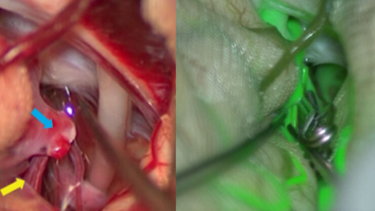
Aneurysm Clipping: Assessing Perforators in Real-time with AR Fluorescence
This article covers two aneurysm clipping cases highlighting the clinical benefits of GLOW800 Augmented Reality Fluorescence application in neurosurgery, based on insights from Prof. Tohru Mizutani,…
Loading...

How to Achieve Brain Tissue Resection with GLOW400 AR
Intraoperative MRI is one form of real-time intraoperative visualization, but if more in-depth visualization to identify a tumor during surgery is wanted, intraoperative fluorescence diagnostics is…
Loading...

组织中的精密空间蛋白质组学信息
尽管可使用基于成像和质谱的方法进行空间蛋白质组学研究,但是图像与单细胞分辨率蛋白丰度测量值的关联仍然是个巨大的挑战。最近引入的一种方法,深层视觉蛋白质组学(DVP),将细胞表型的人工智能图像分析与自动化的单细胞或单核激光显微切割及超高灵敏度的质谱分析结合在了一起。DVP在保留空间背景的同时,将蛋白丰度与复杂的细胞或亚细胞表型关联在一起。
Loading...
![[Translate to chinese:] Multiplexed Cell DIVE imaging of Adult Human Alzheimer’s brain tissue section demonstrating expression of markers specific to astrocytes , microglia , AD-associated markers and immune cells clustered around the β-amyloid plaques [Translate to chinese:] Multiplexed Cell DIVE imaging of Adult Human Alzheimer’s brain tissue section demonstrating expression of markers specific to astrocytes (GFAP, S100B), microglia (TMEM119, IBA1), AD-associated markers (p-Tau217, β-amyloid) and immune cells such as CD11b+, CD163+, CD4+, and HLA-DRA+, clustered around the β-amyloid plaques.](/fileadmin/_processed_/b/b/csm_Alzheimers_brain_tissue_section_showing_astrocytes_microglia_immune_cells_6f24036a9f.jpg)
Spatial Analysis of Neuroimmune Interactions in Alzheimer’s Disease
Alzheimer’s disease (AD) is a complex neurodegenerative disorder characterized by neurofibrillary tangles, β-amyloid plaques, and neuroinflammation. These dysfunctions trigger or are exacerbated by…
Loading...
![[Translate to chinese:] Image of magnetic steel taken with a 100x objective using Kerr microscopy. The magnetic domains in the grains appear in the image with lighter and darker patterns. A few domains are marked with red arrows. [Translate to chinese:] Image of magnetic steel taken with a 100x objective using Kerr microscopy. The magnetic domains in the grains appear in the image with lighter and darker patterns. A few domains are marked with red arrows. Courtesy of Florian Lang-Melzian, Robert Bosch GmbH, Germany.](/fileadmin/_processed_/1/8/csm_Magnetic_steel_100x_objective_Kerr_microscopy_06ead95143.jpg)
使用克尔显微镜快速可视化钢中的磁畴
磁性材料中磁畴与偏振光相互作用后光的旋转,称为克尔效应,使得使用克尔显微镜对磁化样品进行研究成为可能。它可以快速可视化材料表面的磁域。对于用于电气和电子设备的磁性材料(例如钢合金)的高效研发和质量控制,克尔显微镜可以发挥重要作用。本文详细描述了如何使用克尔显微镜对钢合金晶粒中的磁域进行成像。
Loading...
![[Translate to chinese:] According to Dr. Flanagan, the PROvido IVA surgical microscope provides optimal visualization, supporting the needs of tympanoplasty surgery. Image courtesy of Dr. Sean Flanagan. [Translate to chinese:] According to Dr. Flanagan, the PROvido IVA surgical microscope provides optimal visualization, supporting the needs of tympanoplasty surgery. Image courtesy of Dr. Sean Flanagan.](/fileadmin/_processed_/e/e/csm_Tympanoplasty_surgery_with_PROvido_IVA_by_Dr._Flanagan_94faeea788.jpg)
鼓室成形术:优选方法和工具
鼓室成形术用于修复鼓膜穿孔。鼓室成形术有五种类型。第一种鼓室成形术,也称为鼓膜成形术,仅限于鼓膜的修复,而不涉及中耳的其他手术操作。鼓膜成形术是一种常见的但往往被低估的手术。手术方法的选择取决于鼓膜穿孔的位置。良好的可视化是成功修复的关键。
Loading...
![[Translate to chinese:] Intestinal organoids label with FUCCI reporter to follow cell cycle dynamics. Courtesy of Franziska Moos. Liberali lab. FMI Basel (Switzerland). [Translate to chinese:] Intestinal organoids label with FUCCI reporter to follow cell cycle dynamics. Courtesy of Franziska Moos. Liberali lab. FMI Basel (Switzerland).](/fileadmin/_processed_/3/0/csm_Explore-life-events-with-long-term-imaging_a4612dd47d.jpg)
双视野光片显微镜,适用于大型多细胞系统
展示复杂多细胞系统的动态是生物学中的一个基本目标。为了应对在大型时空尺度上进行活体成像的挑战,作者在《自然·方法》杂志上发表的一篇论文中介绍了一种开放式多样本双视野光片显微镜。研究发现,Viventis LS2 Live显微镜在以单细胞分辨率成像大型样本方面取得了显著进展。


