Filter articles
태그
产品
Loading...

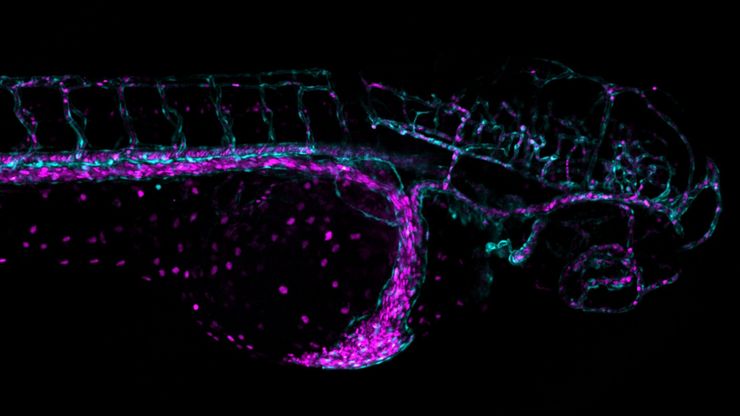
Overcoming Challenges with Microscopy when Imaging Moving Zebrafish Larvae
Zebrafish is a valuable model organism with many beneficial traits. However, imaging a full organism poses challenges as it is not stationary. Here, this case study shows how zebrafish larvae can be…
Loading...
![[Translate to chinese:] Block-face created by automatic trimming under fluorescence. Mammalian cells of interest, stained with CellTrackerTM Green are visualized within the block-face using the UC Enuity. [Translate to chinese:] Block-face created by automatic trimming under fluorescence. Mammalian cells of interest, stained with CellTrackerTM Green are visualized within the block-face using the UC Enuity equipped with the stereo microscope M205 FA. In the background a carbon finder grid in black is visible. All samples in the article are created by Felix Gaedke, PhD, CECAD, Cologne, Germany.](/fileadmin/_processed_/c/c/csm_Block_face_created_by_automatic_trimming_using_fluorescence_527c668077.jpg)
如何在块面中自动获取感兴趣的荧光细胞
本文介绍了使用超薄切片超薄切片机自动修整修块功能,获取树脂块面中带有荧光信号的细胞结构。我们展示了如何使用配置有体视显微镜 M205 FA 的超薄切片超薄切片机 UC Enuity ,来识别感兴趣的荧光细胞,如何自动修整包含细胞的块面,以及如何在切片中观察细胞而无需转移到外部显微镜。
Loading...

A Guide to Spatial Biology
What is spatial biology, and how can researchers leverage its tools to meet the growing demands of biological questions in the post-omics era? This article provides a brief overview of spatial biology…
Loading...

An Introduction to Laser Microdissection
The heterogeneity of histological and biological specimens often requires isolation of specific single cells or cell groups from surrounding tissue before molecular biology analysis can be carried…
Loading...
![[Translate to chinese:] Mouse brain (left) microdissected with a 10x objective (upper right). Inspection of the collection device (lower right). [Translate to chinese:] Mouse brain (left) microdissected with a 10x objective (upper right). Inspection of the collection device (lower right).](/fileadmin/_processed_/f/3/csm_Mouse_brain_microdissected_with_10x_objective_5fbd8963bf.jpg)
激光微切割(LMD)促进的分子生物学分析
使用激光微切割(LMD)提取生物分子、蛋白质、核酸、脂质和染色体,以及提取和操作细胞和组织,可以深入了解基因和蛋白质的功能。它在神经生物学、免疫学、发育生物学、细胞生物学和法医学等多个领域有广泛应用,例如癌症和疾病研究、基因改造、分子病理学和生物学。LMD 也有助于研究蛋白质功能、分子机制及其在转导途径中的相互作用。
Loading...
![[Translate to chinese:] Image of murine dopaminergic neurons which have been marked for laser microdissection (LMD). [Translate to chinese:] Image of murine dopaminergic neurons which have been marked for laser microdissection (LMD).](/fileadmin/_processed_/b/f/csm_Murine_dopaminergic_neurons_marked_for_LMD_d784dbd83f.jpg)
利用激光显微切割(LMD)在空间背景下分离神经元
在阿尔茨海默病之后,帕金森病是第二常见的进行性神经退行性疾病。在首发症状出现之前,中脑中高达70%的多巴胺释放神经元已经死亡。本文描述了如何使用现代激光显微切割(LMD)方法帮助解决帕金森病之谜。研究涉及在空间背景下分离和分析神经元。这些细胞来自帕金森病患者的死后黑质组织样本,以便深入了解该病的分子机制。
Loading...

6英寸晶圆检测显微镜:可靠观察细微高度差异
本文介绍了一种配备自动化和可重复的DIC(微分干涉对比)成像的6英寸晶圆检测显微镜,无论用户的技能水平如何。制造集成电路(IC)芯片和半导体组件需要进行晶圆检测,以验证是否存在影响性能的缺陷。这种检测通常使用光学显微镜进行质量控制、故障分析和研发。为了有效地可视化晶圆上结构之间的小高度差异,可以使用DIC。
Loading...
![[Translate to chinese:] Camera image during auto alignment. The feedback lines indicate if the correct edges in the image are detected. Green: Vertical center line [Translate to chinese:] Camera image during auto alignment. The feedback lines indicate if the correct edges in the image are detected. Green: Vertical center line; Magenta: Upper edge of the light gap; White: Lower edge of the light gap (not visible here, falling together with red line); Red: Knife edge; Blue: Left and right edge of the block face being automatically detected.](/fileadmin/_processed_/d/1/csm_Camera_image_during_auto_alignment_UC_Enuity_5e6b7b9057.jpg)
高质量超薄切片:样品与切片刀自动对齐
超薄切片技术是获取样品切片的最常用方法。在室温条件制备时,将样品小块嵌入环氧树脂中,然后通过修剪去除多余的树脂,并使用玻璃刀或金刚石刀将样品切成厚度为50-100纳米之间的薄片。

![[Translate to chinese:] Section ribbons with increasing section thickness - silver to purple ending in blue sections. [Translate to chinese:] Section ribbons with increasing section thickness - silver to purple ending in blue sections.](/fileadmin/_processed_/7/2/csm_Section_ribbons_with_increasing_section_thickness_d0e10995bf.jpg)
