
Loading...
.")
Spatial Metabolomics: Exploring Tumor Complexity and Therapeutic Insights
In cancer research, it is vital to understand the interaction between tumor cells and their microenvironment, as the tumor microenvironment influences tumor progression significantly. Spatial…
Loading...

Advancing Uterine Regenerative Therapies with Endometrial Organoids
Prof. Kang's group investigates important factors that determine the uterine microenvironment in which embryo insertion and pregnancy are successfully maintained. They are working to develop new…
Loading...

Lipidomics Analysis of Sparse Cells based on Laser Microdissection
Delve into cellular intricacies with high-coverage targeted lipidomics analysis of sparse cells. This advanced method, integrating Laser Microdissection (LMD) and Liquid Chromatography-Mass…
Loading...

Rapidly Visualizing Magnetic Domains in Steel with Kerr Microscopy
The rotation of polarized light after interaction with magnetic domains in a material, known as the Kerr effect, enables the investigation of magnetized samples with Kerr microscopy. It allows rapid…
Loading...
![[Translate to chinese:] AI-based transfection analysis (left) of U2OS cells which were transfected with a fluorescently labelled protein. A fluorescence image of the cells (right) is also shown. The analysis and imaging were performed with Mateo FL.](/fileadmin/_processed_/4/c/csm_AI-based_analysis_of_U2OS_cells_transfected_with_fluorescently_labelled_protein_65d00c857b.jpg "[Translate to chinese:] AI-based transfection analysis (left) of U2OS cells which were transfected with a fluorescently labelled protein. A fluorescence image of the cells (right) is also shown. The analysis and imaging were performed with Mateo FL.")
利用AI实现细胞转染的高效分析
本文探讨了AI(AI)在优化 2D 细胞培养研究中转染效率测量中的关键作用。对于理解细胞机制而言,精确可靠的 2D 细胞培养转染效率测量至关重要。靶向蛋白的高转染效率对于包括活细胞成像和蛋白纯化在内的实验至关重要。手动估计存在不一致性和不可靠性。借助AI的力量,可以实现高效可靠的转染研究。
Loading...
![[Translate to chinese:] Image of confluent cells taken with phase contrast (left) and analyzed for confluency using AI (right).](/fileadmin/_processed_/3/6/csm_Confluent_cells_with_phase_contrast_and_analyzed_for_confluency_using_AI_7c617d0a36.jpg "[Translate to chinese:] Image of confluent cells taken with phase contrast (left) and analyzed for confluency using AI (right).")
通过 AI 汇合度提高 2D 细胞培养的精度
本文解释了如何利用人工智能(AI)进行高效、精确的 2D 细胞培养汇合度评估。准确评估细胞培养的汇合度,即表面积覆盖的百分比,对于可靠的细胞研究至关重要。传统方法使用视觉检查或简单算法,使结果不客观和精确,尤其是对于用于药物发现、组织工程和再生医学的复杂细胞系。利用自动化图像分析和深度学习算法的方法提供更好的精度,并可以增强实验结果。
Loading...
![[Translate to chinese:] Image of murine dopaminergic neurons which have been marked for laser microdissection (LMD).](/fileadmin/_processed_/b/f/csm_Murine_dopaminergic_neurons_marked_for_LMD_cdc0cd9eda.jpg "[Translate to chinese:] Image of murine dopaminergic neurons which have been marked for laser microdissection (LMD).")
利用激光显微切割(LMD)在空间背景下分离神经元
在阿尔茨海默病之后,帕金森病是第二常见的进行性神经退行性疾病。在首发症状出现之前,中脑中高达70%的多巴胺释放神经元已经死亡。本文描述了如何使用现代激光显微切割(LMD)方法帮助解决帕金森病之谜。研究涉及在空间背景下分离和分析神经元。这些细胞来自帕金森病患者的死后黑质组织样本,以便深入了解该病的分子机制。
Loading...
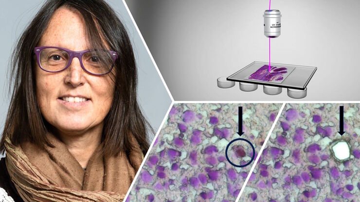
激光显微切割技术如何助力神经科学研究取得开创性进展?
玛尔塔·帕特林尼博士,卡罗林斯卡学院的高级科学家,分享了她在成人人类神经发生开创性研究中使用激光显微切割(LMD)的经验,并提供了关于LMD在空间蛋白质组学和精准医学中未来应用潜力的个人见解。
Loading...
![[Translate to chinese:] How the M822 microscope enhances surgical precision in eye surgeries - Insights from Dr. Dhami. Image courtesy of Dr. Abhinav Dhami.](/fileadmin/_processed_/d/3/csm_Dr_Abhinav_Dhami_with_M822_28434efe21.jpg "[Translate to chinese:] How the M822 microscope enhances surgical precision in eye surgeries - Insights from Dr. Dhami. Image courtesy of Dr. Abhinav Dhami.")
听听Dhami 医生关于购买眼科显微镜的专业见解
在本文中,了解来自印度北部的眼科手术顾问Abhinav Dhami医生如何使用Leica Microsystems的M822眼科显微镜来提高他的手术精确度,以及哪些关键特性使他决定购买这款眼科显微镜用于他的实践。