22
–
23
Apr
2025
第四届半导体封装检测及失效分析技术进展网络研讨会
China
•
Webinar
22
Apr
2025
共聚焦荧光寿命成像功能在植物相关研究中的应用
China
•
Webinar
Filter articles
标签
产品
Loading...
.")
您的 3D 类器官成像和分析工作流程效率如何?
类器官模型已经改变了生命科学研究,但优化图像分析协议仍然是一个关键挑战。本次网络研讨会探讨了类器官研究的简化工作流程,首先是实时的三维细胞培养检查,接下来是高速、高分辨率的三维成像,生成清晰的图像和更纯净的数据,以便对生长速率、细胞迁移和三维细胞相互作用等参数进行准确地人工智能分割和量化,从而实现更深入的洞察。
Loading...

细胞活成像的纳米级扩展
新的STED显微技术方法——TauSTED Xtend,使得在纳米级别下对活体完整样本进行扩展多色成像成为可能。通过结合空间和寿命信息,TauSTED Xtend提供了额外一层信息,允许在极低的光剂量下分辨小细节并在整体结构中解析它们。
Loading...
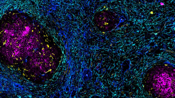
加速不同组织多重成像的发现
组织的多重成像对于肿瘤-免疫相互作用的研究以及人类细胞图谱等发现工作越来越重要。 欢迎加入我们的演讲,Andrea J. Radtke 博士解释了如何使用迭代漂白扩展多重性 (IBEX) 绘制组织图谱,并讨论了用于多重成像的广泛社区资源。
Loading...

Transforming Multiplexed 2D Data into Spatial Insights Guided by AI
Aivia 13 handles large 2D images and enables researchers to obtain deep insights into microenvironment surrounding their phenotypes with millions of detected objects and automatic clustering up to 30…

Loading...

共聚焦多色成像在癌症研究和免疫学中的潜力
在本次网络研讨会上,来自莫纳什制药科学研究所的CameronNowell和他的同事将分享他们在多重成像方面的经验,以及他们通过巧妙的共聚焦成像采集和利用FLIM等其他多重成像模式所取得的成果。
