Webinars

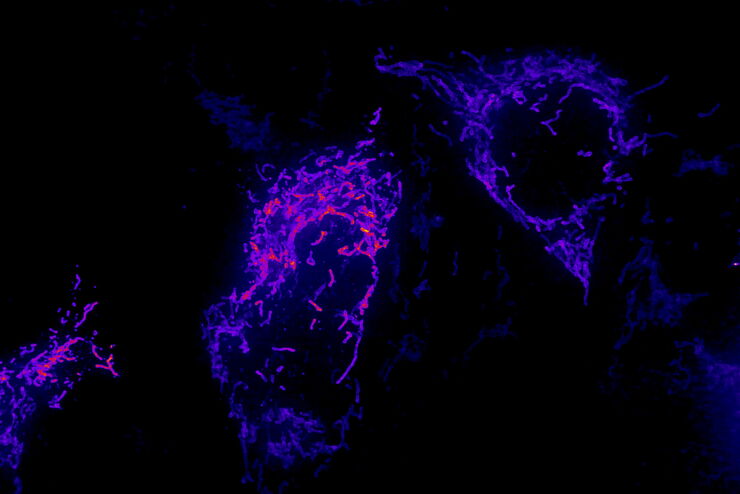
Image of fixed U2OS cell expressing mEmerald-Tomm20 denoised using a 3D RCAN model trained with matching low and high SNR image pairs acquired on an iSIM system.

人工智能显微图像分析-介绍

人工智能引领的显微图像分析和可视化是用于数据驱动型科学发现的一项强大工具。人工智能技术可以帮助研究人员应对具有挑战性的成像应用,让他们能够从图像中获取更多的信息。
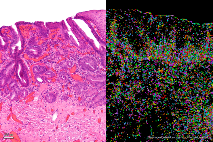
H&E stained micrograph of an intramucosal esophageal adenocarcinoma (left) enhanced with Aivia’s Pixel Classifier (right)

简化癌症生物学图像分析工作流

随着癌症生物学数据集的不断增长,显微图像分割和定量也越来越具挑战性,研究人员被迫在分析工作中耗费大量的时间。
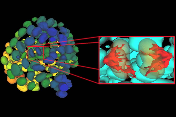
Single timepoint of a time-lapse recording of mammary epithelial micro spheroid cultured in 3D highlighting individual mitotic events

在不同尺度下观察复杂的细胞相互作用

细胞间的相互作用很难观察,其中涉及的目标检测和关系衡量尤为棘手。没有简单易用的目标检测及其关系测量方法,很难观察到细胞间的相互作用。
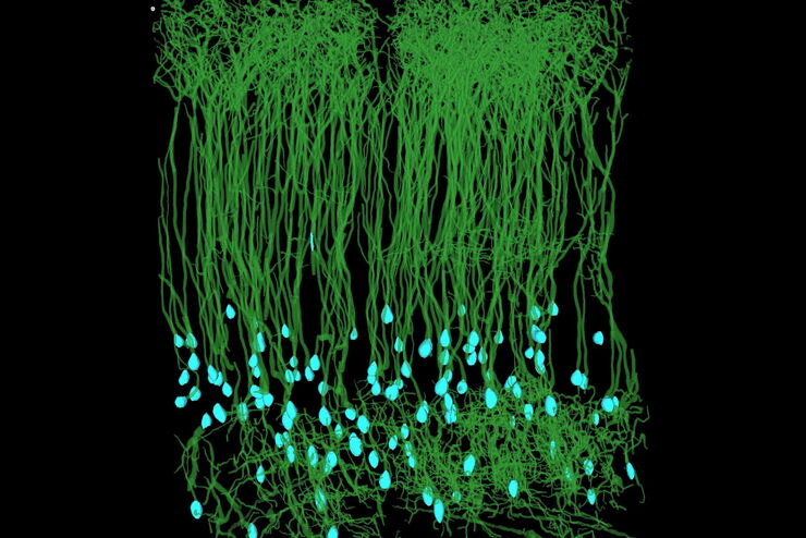
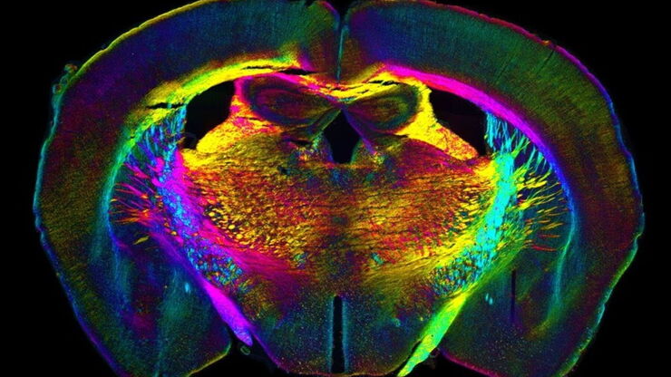
Aivia_Neuroscience-VBE comparison mouse-1_traced_ROI

自动化加速神经元图像分析

复杂神经投射的检测能力主要取决于大规模神经元网络的精确重建。神经科学研究中的大多数数据析取方法都非常耗时和易错,进而导致进度延误和错误。在本次研讨会中,Aivia将演示如何利用自动化技术提升图像分析工作流的效率
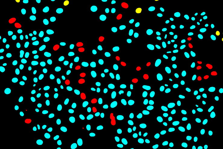
Separation of cells based on their tracking status: A colourised binary mask of a time-lapse microscopy field of view of medium confluency with individual cells highlighted as survivors if they can be tracked since the initial movie frame (cyan), incomers if they migrated into the field of view throughout the movie (yellow) or mistracks if an error occurred in the automated trajectory reconstruction (red).

使用深度学习技术追踪单细胞

人工智能解决方案在显微镜领域的应用不断拓展。从自动化目标分类到虚拟染色,机器学习和深度学习技术在帮助显微镜学家简化分析工作的同时,也在持续推动科学技术领域的突破。
Analysis of anatomy and axon orientation of an adult mouse brain tissue with QLIPP.

通过光学属性了解细胞结构

在过去3年中,显微学家开始在广泛的应用中使用人工智能解决方案,包括图像采集优化(智能显微镜)、目标分类、图像分类、分割、还原、超分辨率和虚拟染色。
Scroll to top