Webinars

最新市场活动资讯

全部活动

16
22 Apr 2025
Image: Human stem cell-derived mid brain organoids. Courtesy of Dr Tanya Singh, University of Oxford.

Unlocking the Secrets of Organoid Models in Biomedical Research

Get ready to delve deeper into the world of organoids and 3D models, which are essential tools for advancing our understanding of human health. Navigating these complex structures and obtaining clear…
Multicolor fixed STED image. Inner ear section, mouse, TauSTED Xtend 589 on AF488 and TauSTED Xtend 775 on AF633-Phalloidin. Sample courtesy of Dennis Derstrof, Klinik für Hals-, Nasen und Ohrenheilkunde, Universität Marburg & Prof. Dr. Dominik Oliver aus dem Institut für Physiologie und Pathophysiologie, Abteilung für Neurophysiologie, Universität Marburg.

细胞活成像的纳米级扩展

新的STED显微技术方法——TauSTED Xtend,使得在纳米级别下对活体完整样本进行扩展多色成像成为可能。通过结合空间和寿命信息,TauSTED Xtend提供了额外一层信息,允许在极低的光剂量下分辨小细节并在整体结构中解析它们。
In vivo imaging of a mouse pial and cortical vasculature through a glass window (ROSAmT/mG::Pdgfb-CreERT2 mouse meningeal and cortical visualization following tamoxifen induction and craniotomy). Courtesy: Thomas Mathivet, PhD

神经血管病理学之窗

探索先天性免疫如何在神经血管病变后持续产生有害影响,以及能够对这些事件进行纵向研究的技术发展。
Lifetime-based multiplexing in live cells using TauSeparation. Mammalian cells expressing LifeAct-GFP (ibidi GmbH) and labelled with MitoTracker Green. Acquisition with one detector, intensity information shown in grey. The two markers can be separated using lifetime information: LifeAct-GFP (cyan), MitoTracker Green (magenta). Image acquired with STELLARIS 5.

可重复性、协作和新成像技术的力量

在本次网络研讨会上,您将了解到影响显微镜可重复性的因素,有哪些资源和举措可用于改善显微镜教育并提高其严谨性和可重复性以及研究人员、成像科学家和显微镜供应商之间的合作如何推动创新和采用新技术。

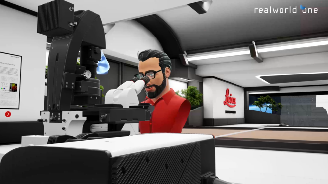
Virtual Reality Showcase for STELLARIS Confocal Microscopy Platform

In this webinar, you will discover how to perform 10-color acquisition using a confocal microscope. The challenges of imaged-based approaches to identify skin immune cells. A new pipeline to assess…

免疫细胞在组织样品中的共聚焦成像

在本次网络研讨会中,您将探索如何使用共聚焦显微镜对组织样品进行10色成像,并了解这一技术如何有助于评估皮肤免疫状况。
Scroll to top