
Filter articles
标签
产品
Loading...

Coherent Raman Scattering Microscopy Publication List
CRS (Coherent Raman Scattering) microscopy is an umbrella term for label-free methods that image biological structures by exploiting the characteristic, intrinsic vibrational contrast of their…

Loading...
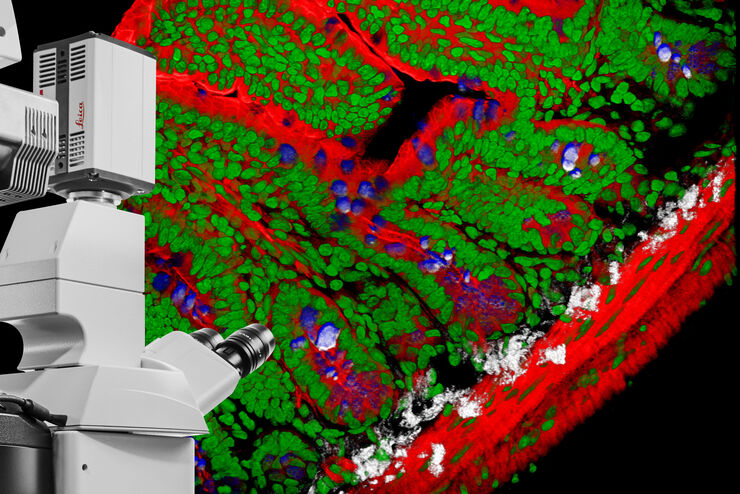
DIVE Multiphoton Microscope Image Gallery
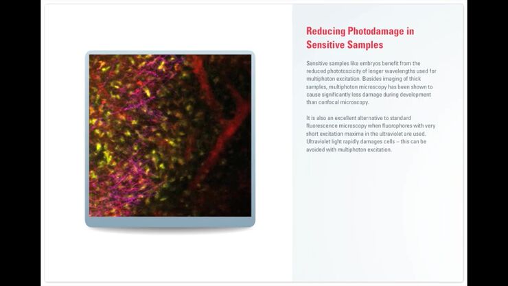
Today’s life science research focusses on complex biological processes, such as the causes of cancer and other human diseases. A deep look into tissues and living specimens is vital to understanding…

Loading...

不可能的任务:可调颜色用于非扫描检测
徕卡显微系统的 4Tune 探测器是 SP8 DIVE 深度体内探测器的关键组件,提供具有非扫描检测的光谱可调图像记录,是多参数多光子显微镜的创新解决方案。
Loading...

Laser Beam Shaping for Multicolor Multiphoton Microscopy
Multiphoton Microscopy is one of the current hot topics in life science research. The new Leica TCS SP8 DIVE from Leica Microsystems presents a series of beneficial new innovations, including a freely…
Loading...
高分辨率共聚焦显微镜的 BABB 清洗和成像
Multipohoton microscopy experiment using Leica TCS SP8 MP and Leica 20x/0.95 NA BABB immersion objective.
Understanding kidney microanatomy is key to detecting and identifying early events in kidney…
Loading...

CARS 相干反斯托克斯拉曼散射显微镜: 分子特征振动对比成像
相干反斯托克斯拉曼散射(CARS)显微技术是一种根据分子振动特征生成图像的技术。这种成像方法不需要标记,但可以从一系列重要的生物分子化合物中获得特定的分子信息。
