显微镜知识库

显微镜知识库

徕卡显微系统的知识库提供有关显微镜学科的科学研究和教学材料。内容旨在对显微镜初学者、有经验的显微镜操作实践者和使用显微镜的科学家在他们的日常工作和实验有所帮助。这里有探索交互式教程和应用笔记,你可以找到你需要的显微镜的基础知识以及前沿技术——快来加入徕卡显微知识社区,分享您的专业知识!
Transfection using the Uncommon Bio reprogramming system. Image acquired using the THUNDER Imager 3D Cell Culture with THUNDER Large Volume Computational Clearing (LVCC) applied. Image courtesy of Samuel East, Uncommon Bio.

利用新型可扩展的干细胞培养设计未来

具有远见卓识的生物技术初创企业 Uncommon Bio 正在应对世界上最大的健康挑战之一:食品可持续性。在这次网络研讨会上,干细胞科学家塞缪尔-伊斯特(Samuel East)将展示他们如何使细胞农业的干细胞培养基既安全又经济可行。了解他们如何将培养基成本降低 1000 倍,并开发出不含动物成分、食品安全的 iPSC 培养基。
3D culture of ovarian cancer cells imaged using the confocal mode of Mica.

Mica: 助力伦敦帝国学院开展跨学科科研研究

这篇访谈重点介绍了伦敦帝国学院的 Mica 所产生的变革性影响。科学家们解释了Mica如何改变了游戏规则,扩大了研究的可能性,促进了跨学科合作。他们解释了使用 Mica 进行详细的活细胞成像如何提供更有意义的信息,使科学家始终站在研究的最前沿。研究小组预计,Mica将继续开辟新的研究途径,包括研究微流体技术和其他先进应用。
Developing embryos of different species at different stages during the elongation of their posterior body axis, from left to right in developmental time. The labelled regions in red depict a region of undifferentiated cells called the tailbud, with the corresponding region generated from that tissue shaded in grey. Upper row: lamprey; middle row: catshark; bottom row, zebrafish. This figure has been adapted from the following publication: Steventon, B., Duarte, F., Lagadec, R., Mazan, S., Nicolas, J.-F., & Hirsinger, E. (2016). Species tailoured contribution of volumetric growth and tissue convergence to posterior body elongation in vertebrates. Development, 2016. 143(10):1732-41

How to Study Gene Regulatory Networks in Embryonic Development

Join Dr. Andrea Boni by attending this on-demand webinar to explore how light-sheet microscopy revolutionizes developmental biology. This advanced imaging technique allows for high-speed, volumetric…
Salmonella biofilms 3D render

探索微生物世界:三维食品基质中的空间相互作用

Micalis 研究所是与 INRAE、AgroParisTech 和巴黎萨克雷大学合作的联合研究单位。其使命是开发食品微生物学领域的创新研究,以促进健康。在这一系列视频中,Micalis…
Dapi – Nucleus, GFP – Plasma Membrane, Thickness 100µm, 63x objektive, 469 Z planes, 2 channels, THUNDER Imager 3D Cell Culture. Courtesy M.Sc. Dana Krauß, Medical University of Vienna (Austria).

您的 3D 类器官成像和分析工作流程效率如何?

类器官模型已经改变了生命科学研究,但优化图像分析协议仍然是一个关键挑战。本次网络研讨会探讨了类器官研究的简化工作流程,首先是实时的三维细胞培养检查,接下来是高速、高分辨率的三维成像,生成清晰的图像和更纯净的数据,以便对生长速率、细胞迁移和三维细胞相互作用等参数进行准确地人工智能分割和量化,从而实现更深入的洞察。
40x magnification of organoids cluster taken on Mateo TL.Cell type: esophageal squamous carcinoma; scale  bar 15µm. Courtesy of bioGenous, China.

克服类器官三维细胞培养中的观察挑战

类器官在细胞生物学和药物发现中至关重要,因为它们能够模拟体内细胞的复杂性和结构,有助于癌症等微环境至关重要的疾病研究。类器官可根据患者的基因型进行定制,这也有助于个性化医学研究。
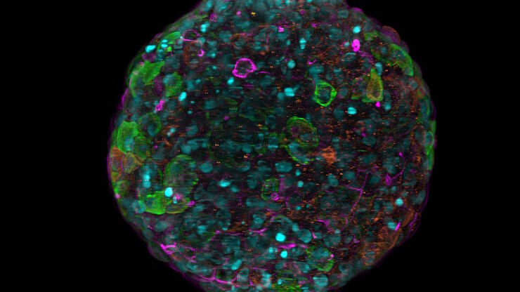
Spheroid stained with Cyan: Dapi nuclear countertain; Green AF488 Involucrin; Orange AF55 Phalloidin Actin; Magenta AF647 CK14.

基于人工智能的表型药物筛查解决方案

本次网络研讨会将全面介绍使用三维细胞培养进行表型药物筛选所遇到的问题、可能的解决方案及规划与执行策略。
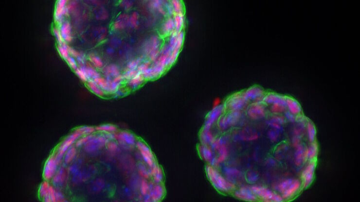
Murine esophageal organoids (DAPI, Integrin26-AF 488, SOX2-AF568) imaged with the THUNDER Imager 3D Cell Culture. Courtesy of Dr. F.T. Arroso Martins, Tamere University, Finland.

如何深入了解类器官和细胞球模型

在本电子书中,您将了解3D细胞培养模型(如类器官和细胞球)成像的关键注意事项。探索创新型显微镜解决方案,来实时记录类器官和细胞球的动态成像过程。
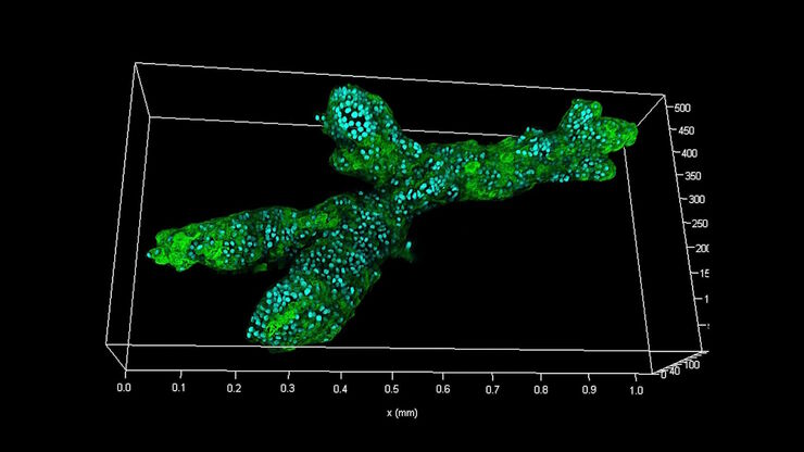
Branched organoid growing in collagen where the Nuclei are labeled blue. To detect the mechanosignaling process, the YAP1 is labeled green.

检查癌症类器官的发展进程

德国慕尼黑工业大学的Andreas Bausch实验室研究细胞和生物体中不同结构和功能形成的细胞和生物物理机制。他的团队设计了新的策略、方法和分析工具,以量化微米和纳米等级的发展机制和动态过程。关键研究领域包括干细胞和类器官,从乳腺类器官到胰腺癌类器官,以更好地了解疾病模型。
Scroll to top