26
Feb
2025
Designing the Future with Novel Stem Cell Culture
United Kingdom
•
Webinar
26
Feb
2025
Designing the Future: Novel Stem Cell Culture and RNA Tech
Singapore
•
Webinar
Filter articles
标签
产品
Loading...
![[Translate to chinese:] UC Enuity](/fileadmin/_processed_/e/9/csm_UC_Enuity_Detail_Header_411ca2e94e.jpg "UC Enuity")
通过自动切片改善您的超薄切片工作流程
在不断发展的电镜样品制备领域,保持领先地位至关重要。这个网络研讨会提供了关于超薄切片最新进展的重要见解,这些进展可以显著增强您实验室的能力。
Loading...
![[Translate to chinese:] The role of extracellular signalling mechanisms in the correct development of the human brain](/fileadmin/_processed_/a/e/csm_The_role_of_extracellular_signalling_mechanisms_in_the_correct_development_of_the_human_brain_6b9e3b80f0.jpg "[Translate to chinese:] The role of extracellular signalling mechanisms in the correct development of the human brain")
在神经发育过程中,细胞是如何相互交流的?
细胞间通信是大脑发育过程中一个必不可少的过程,它受到多种因素的影响,包括细胞的形态、粘附分子、局部细胞外基质和分泌囊泡。在本次网络研讨会上,您将了解到对这些机制更深入的理解是如何推动对神经发育障碍的理解的。
Loading...
![[Translate to chinese:] Multicolor fixed STED image. Inner ear section, mouse, TauSTED Xtend 589 on AF488 and TauSTED Xtend 775 on AF633-Phalloidin. Sample courtesy of Dennis Derstrof, Klinik für Hals-, Nasen und Ohrenheilkunde, Universität Marburg & Prof. Dr. Dominik Oliver aus dem Institut für Physiologie und Pathophysiologie, Abteilung für Neurophysiologie, Universität Marburg.](/fileadmin/_processed_/c/a/csm_Inner_ear_section_mouse_multicolor_fixed_TauSTED_Xtend_09ebb898fd.jpg "[Translate to chinese:] Multicolor fixed STED image. Inner ear section, mouse, TauSTED Xtend 589 on AF488 and TauSTED Xtend 775 on AF633-Phalloidin.")
细胞活成像的纳米级扩展
新的STED显微技术方法——TauSTED Xtend,使得在纳米级别下对活体完整样本进行扩展多色成像成为可能。通过结合空间和寿命信息,TauSTED Xtend提供了额外一层信息,允许在极低的光剂量下分辨小细节并在整体结构中解析它们。
Loading...
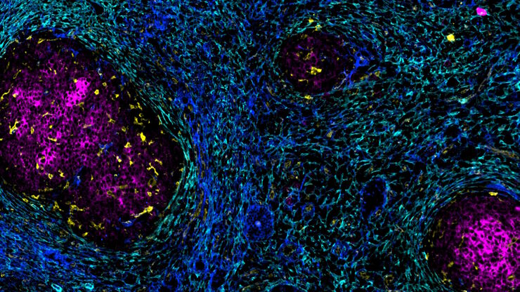
加速不同组织多重成像的发现
组织的多重成像对于肿瘤-免疫相互作用的研究以及人类细胞图谱等发现工作越来越重要。 欢迎加入我们的演讲,Andrea J. Radtke 博士解释了如何使用迭代漂白扩展多重性 (IBEX) 绘制组织图谱,并讨论了用于多重成像的广泛社区资源。
Loading...

Transforming Multiplexed 2D Data into Spatial Insights Guided by AI
Aivia 13 handles large 2D images and enables researchers to obtain deep insights into microenvironment surrounding their phenotypes with millions of detected objects and automatic clustering up to 30…
![[Translate to chinese:] Spheroid stained with Cyan: Dapi nuclear countertain; Green AF488 Involucrin; Orange AF55 Phalloidin Actin; Magenta AF647 CK14.](/fileadmin/_processed_/6/8/csm_Spheroid_stained_with_Cyan_4color_overlay_78dff87b83.jpg "[Translate to chinese:] Spheroid stained with Cyan: Dapi nuclear countertain; Green AF488 Involucrin; Orange AF55 Phalloidin Actin; Magenta AF647 CK14.")
