Filter articles
标签
产品
Loading...
![[Translate to chinese:] Dr. Ozana Moraru shares two primary open-angle glaucoma cases in which trabeculectomy bleb needling was performed using the Leica M844 microscope with EnFocus intraoperative OCT. Image courtesy of Dr. Ozana Moraru. [Translate to chinese:] Dr. Ozana Moraru shares two primary open-angle glaucoma cases in which trabeculectomy bleb needling was performed using the Leica M844 microscope with EnFocus intraoperative OCT. Image courtesy of Dr. Ozana Moraru.](/fileadmin/_processed_/1/0/csm_Trabeculectomy_bleb_needling_performed_with_Leica_M844_microscope_with_EnFocus_intraoperative_OCT_14e8587e0d.jpg)
术中光学显像如何帮助青光眼手术获得更多洞察力
青光眼是导致全球失明的主要原因之一。青光眼手术可以延缓疾病的发展。在青光眼手术过程中,术中光学相干断层扫描(OCT)的使用为眼科外科医生提供了更佳的可视化效果,让他们更深入地了解表面下组织对手术操作的反应。 莫拉鲁博士通过两个原发性开角型青光眼(POAG)的临床病例强调了它的价值。
Loading...

眼科: 复杂白内障手术中的可视化
白内障手术是最常见的眼科手术。为了满足白内障手术的需要,Ozana Moraru 博士使用了 Leica Microsystems 的 M844 显微镜和 EnFocus 术中光学相干断层扫描 (OCT) 以及 3D 可视化系统。在本案例研究中,她介绍了术中光学相干断层扫描如何为标准和复杂的白内障手术病例提供有用信息。
Loading...
![[Translate to chinese:] Fundus photo depicting epiretinal membrane with lamellar macular hole. Image courtesy of Robert A. Sisk, MD, FACS, Cincinnati Eye Institute. [Translate to chinese:] Fundus photo depicting epiretinal membrane with lamellar macular hole. Image courtesy of Robert A. Sisk, MD, FACS, Cincinnati Eye Institute.](/fileadmin/_processed_/e/8/csm_Epiretinal-membrane-with-lamellar-macular-hole_0d86f1b2dd.jpg)
使用光学相干断层扫描改善黄斑孔手术
黄斑裂孔是一种罕见的眼疾,会导致中心视力模糊,影响日常活动。黄斑裂孔通常是由黄斑被牵拉或拉伸而引起的开口,最常见的原因通常是年龄相关的眼部变化。然而,在本病例研究中,Robert A. Sisk博士(医学博士,FACS)介绍了一个小儿眼科病例,其中术中光学相干断层扫描(OCT)为他的手术提供了额外的见解。
Loading...
![[Translate to chinese:] Augmented Reality fluorescence supports each step of neurovascular surgery procedures. Image courtesy of Dr. Christof Renner. [Translate to chinese:] Augmented Reality fluorescence supports each step of neurovascular surgery procedures. Image courtesy of Dr. Christof Renner.](/fileadmin/_processed_/4/f/csm_Augmented_Reality_fluorescence_supports_neurovascular_surgery_procedures_bb2c4277c8.jpg)
AR荧光在神经血管手术中的应用
术中血管造影在神经血管手术中发挥着至关重要的作用。在徕卡2021神经可视化峰会期间,Christof Renner博士在独家网络研讨会上展示了精选临床病例,并分享了使用GLOW800增强现实荧光技术的经验。
Loading...
![[Translate to chinese:] Corneal transplantation. Image courtesy of Mr. David Anderson. [Translate to chinese:] Corneal transplantation. Image courtesy of Mr. David Anderson.](/fileadmin/_processed_/e/e/csm_Corneal_transplantation_2beefcbeb9.jpg)
眼科案例分析:角膜移植
内皮角膜移植术是一种现代角膜移植技术。角膜移植手术有多种手术方法,包括角膜后弹力膜撕除角膜内皮移植术(DSEK)和角膜后弹力膜内皮移植术(DMEK)。这些方法在植入供体组织的数量上有所不同。
Loading...
![[Translate to chinese:] PDAC Multiplexed imaging of CST panels enables an examination of immune cell components in pancreatic ductal adenocarcinoma (IPDAC) tissue on a single slide. [Translate to chinese:] PDAC Multiplexed imaging of CST panels enables an examination of immune cell components in pancreatic ductal adenocarcinoma (IPDAC) tissue on a single slide.](/fileadmin/_processed_/a/d/csm_Pancreatic_ductal_adenocarcinoma_tissue_d8790cf699.jpg)
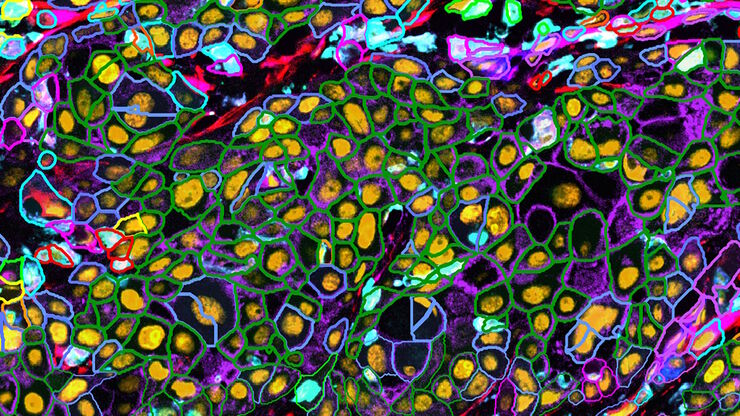
表征肿瘤环境以揭示洞察和空间分辨率
肿瘤环境的表征可以为癌症进展和潜在治疗靶点提供更深入的见解。我们已经使用来自Cell Signaling Technology(CST)的各种IHC验证抗体,在胰腺癌的Cell DIVE研究中验证了30多种偶联抗体。

![[Translate to chinese:] Prof. Nikolaos Bechrakis uses the Proveo 8 ceiling mounted microscope with EnFocus intraoperative OCT. Images provided by Prof. Nikolaos Bechrakis. [Translate to chinese:] Prof. Nikolaos Bechrakis uses the Proveo 8 ceiling mounted microscope with EnFocus intraoperative OCT. Images provided by Prof. Nikolaos Bechrakis.](/fileadmin/_processed_/7/3/csm_Prof_Bechrakis_uses_Proveo_8_ceiling_mounted_microscope_with_EnFocus_intraoperative_OCT_0d79cea5a2.jpg)
![[Translate to chinese:] Keratoplasty of pathologic cornea [Translate to chinese:] Keratoplasty of pathologic cornea](/fileadmin/_processed_/6/d/csm_Pathologic_cornea_633aed9eab.jpg)

