
Filter articles
标签
产品
Loading...
 applied. Image courtesy of Samuel East, Uncommon Bio.")
Designing the Future with Stem Cell and RNA Technology
Visionary biotech start-up Uncommon Bio is tackling one of the world’s biggest health challenges: food sustainability. In this webinar, Stem Cell Scientist Samuel East will show how they use RNA…
Loading...

Revealing Neuronal Migration’s Molecular Secrets
Different approaches can be used to investigate neuronal migration to their niche in the developing brain. In this webinar, experts from The University of Oxford present the microscopy tools and…
Loading...
.")
您的 3D 类器官成像和分析工作流程效率如何?
类器官模型已经改变了生命科学研究,但优化图像分析协议仍然是一个关键挑战。本次网络研讨会探讨了类器官研究的简化工作流程,首先是实时的三维细胞培养检查,接下来是高速、高分辨率的三维成像,生成清晰的图像和更纯净的数据,以便对生长速率、细胞迁移和三维细胞相互作用等参数进行准确地人工智能分割和量化,从而实现更深入的洞察。
![[Translate to chinese:] Murine esophageal organoids (DAPI, Integrin26-AF 488, SOX2-AF568) imaged with the THUNDER Imager 3D Cell Culture. Courtesy of Dr. F.T. Arroso Martins, Tamere University, Finland.](/fileadmin/_processed_/f/f/csm_THUNDER_Imager_3D_Cell_Culture_Murine-esophageal-organoid_LVCC_299fe0ce61.jpg "[Translate to chinese:] Murine esophageal organoids (DAPI, Integrin26-AF 488, SOX2-AF568) imaged with the THUNDER Imager 3D Cell Culture. Courtesy of Dr. F.T. Arroso Martins, Tamere University, Finland.")
Loading...
![[Translate to chinese:] Brain organoid section (DAPI) acquired using THUNDER Imager Live Cell. Image courtesy of Janina Kaspar and Irene Santisteban, Schäfer Lab, TUM.](/fileadmin/_processed_/2/7/csm_Tilescan_of_brain_organoid_section_364be9e906.jpg "[Translate to chinese:] Brain organoid section (DAPI) acquired using THUNDER Imager Live Cell. Image courtesy of Janina Kaspar and Irene Santisteban, Schäfer Lab, TUM.")
研究大脑健康的成像类器官模型
小胶质细胞是特化的脑驻留免疫细胞,在大脑发育、平衡和疾病中发挥着至关重要的作用。然而,到目前为止,模拟人脑环境与小胶质细胞之间相互作用的能力还非常有限。
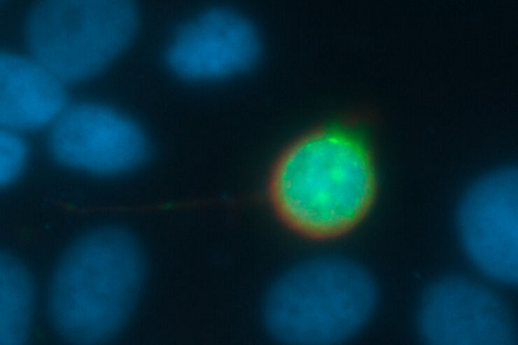
![[Translate to chinese:] Mouse cortical neurons. Transgenic GFP (green). Image courtesy of Prof. Hui Guo, School of Life Sciences, Central South University, China](/fileadmin/_processed_/2/a/csm_THUNDER_Imager_Mouse_cortical_neuron_1fa1718d8f.jpg "[Translate to chinese:] Mouse cortical neurons. Transgenic GFP (green). Image courtesy of Prof. Hui Guo, School of Life Sciences, Central South University, China")
Loading...
![[Translate to chinese:] Microscopy for neuroscience research](/fileadmin/_processed_/9/5/csm_Microscopy_for_neuroscience_research_6a48c90764.jpg "[Translate to chinese:] Microscopy for neuroscience research")
神经科学显微镜面临哪些挑战?
显微镜是神经科学研究领域的强大工具。不过,当涉及到对神经过程进行成像以及使用不同的样品类型(例如厚神经组织或脑类器官)时,科研人员可能会面临到很多挑战。这本30页的电子书包含众多真实的案例,以讨论我们最常见到的一些挑战,同时展示了如何使用THUNDER 成像技术克服这些挑战。
![[Translate to chinese:] Cancer cells](/fileadmin/_processed_/6/b/csm_Cancer_cells_f5e991d1c9.jpg "[Translate to chinese:] Cancer cells")
Loading...
超越反卷积
宽场荧光显微镜通常用于视觉呈现生命科学样本中的结构并获取重要信息。利用荧光蛋白或染料,以高度特异性的方式标记离散的样本部分。为了充分了解某种结构,可能需要以三维方式呈现,但这会对使用显微镜带来某些挑战。
