
Filter articles
标签
产品
Loading...
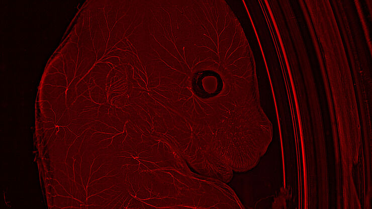
模式生物研究
模式生物是研究人员用来研究特定生物学过程的物种。 它们具有与人类相似的遗传特征,通常用于遗传学、发育生物学和神经科学等研究领域。 选择模式生物的原因通常是它们在实验室环境中易于保持和繁殖、生成周期短,或能够产生突变体来研究某些性状或疾病。
Loading...

Overcoming Challenges with Microscopy when Imaging Moving Zebrafish Larvae
Zebrafish is a valuable model organism with many beneficial traits. However, imaging a full organism poses challenges as it is not stationary. Here, this case study shows how zebrafish larvae can be…
Loading...

How to Study Gene Regulatory Networks in Embryonic Development
Join Dr. Andrea Boni by attending this on-demand webinar to explore how light-sheet microscopy revolutionizes developmental biology. This advanced imaging technique allows for high-speed, volumetric…
Loading...
![[Translate to chinese:] Mouse brain (left) microdissected with a 10x objective (upper right). Inspection of the collection device (lower right).](/fileadmin/_processed_/f/3/csm_Mouse_brain_microdissected_with_10x_objective_5fbd8963bf.jpg "[Translate to chinese:] Mouse brain (left) microdissected with a 10x objective (upper right). Inspection of the collection device (lower right).")
激光微切割(LMD)促进的分子生物学分析
使用激光微切割(LMD)提取生物分子、蛋白质、核酸、脂质和染色体,以及提取和操作细胞和组织,可以深入了解基因和蛋白质的功能。它在神经生物学、免疫学、发育生物学、细胞生物学和法医学等多个领域有广泛应用,例如癌症和疾病研究、基因改造、分子病理学和生物学。LMD 也有助于研究蛋白质功能、分子机制及其在转导途径中的相互作用。
Loading...
![[Translate to chinese:] Image of murine dopaminergic neurons which have been marked for laser microdissection (LMD).](/fileadmin/_processed_/b/f/csm_Murine_dopaminergic_neurons_marked_for_LMD_d784dbd83f.jpg "[Translate to chinese:] Image of murine dopaminergic neurons which have been marked for laser microdissection (LMD).")
利用激光显微切割(LMD)在空间背景下分离神经元
在阿尔茨海默病之后,帕金森病是第二常见的进行性神经退行性疾病。在首发症状出现之前,中脑中高达70%的多巴胺释放神经元已经死亡。本文描述了如何使用现代激光显微切割(LMD)方法帮助解决帕金森病之谜。研究涉及在空间背景下分离和分析神经元。这些细胞来自帕金森病患者的死后黑质组织样本,以便深入了解该病的分子机制。
Loading...

激光显微切割技术用于组织和细胞分离的协议 - 免费下载电子书
激光显微切割(LMD,也称为激光捕获显微切割或LCM)使用户能够分离特定的单个细胞或整个组织区域,甚至亚细胞结构如染色体。纯化的组织和细胞可用于下游的RNA、DNA和蛋白质组工作流程。
Loading...
![[Translate to chinese:] The role of extracellular signalling mechanisms in the correct development of the human brain](/fileadmin/_processed_/a/e/csm_The_role_of_extracellular_signalling_mechanisms_in_the_correct_development_of_the_human_brain_6b9e3b80f0.jpg "[Translate to chinese:] The role of extracellular signalling mechanisms in the correct development of the human brain")
在神经发育过程中,细胞是如何相互交流的?
细胞间通信是大脑发育过程中一个必不可少的过程,它受到多种因素的影响,包括细胞的形态、粘附分子、局部细胞外基质和分泌囊泡。在本次网络研讨会上,您将了解到对这些机制更深入的理解是如何推动对神经发育障碍的理解的。
![[Translate to chinese:] In vivo imaging of a mouse pial and cortical vasculature through a glass window (ROSAmT/mG::Pdgfb-CreERT2 mouse meningeal and cortical visualization following tamoxifen induction and craniotomy). Courtesy: Thomas Mathivet, PhD](/fileadmin/_processed_/6/3/csm_Mouse_pial_and_cortical_vasculature_d3e7a9948c.jpg "[Translate to chinese:] In vivo imaging of a mouse pial and cortical vasculature through a glass window (ROSAmT/mG::Pdgfb-CreERT2 mouse meningeal and cortical visualization following tamoxifen induction and craniotomy). Courtesy: Thomas Mathivet, PhD")
![[Translate to chinese:] Mouse cortical neurons. Transgenic GFP (green). Image courtesy of Prof. Hui Guo, School of Life Sciences, Central South University, China](/fileadmin/_processed_/2/a/csm_THUNDER_Imager_Mouse_cortical_neuron_1fa1718d8f.jpg "[Translate to chinese:] Mouse cortical neurons. Transgenic GFP (green). Image courtesy of Prof. Hui Guo, School of Life Sciences, Central South University, China")
