
Filter articles
标签
产品
Loading...

Virtual Reality Showcase for STELLARIS Confocal Microscopy Platform
In this webinar, you will discover how to perform 10-color acquisition using a confocal microscope. The challenges of imaged-based approaches to identify skin immune cells. A new pipeline to assess…
Loading...
通过非拟合且简便的 FRET-FLIM 方法可视化蛋白质 - 蛋白质相互作用
了解活细胞中的分子相互作用对于解读大多数细胞功能背后的分子机制至关重要。研究蛋白质-蛋白质相互作用的金标准是福斯特共振能量转移(FRET)。尽管有几种方法可以在生物样品中证明FRET,但使用荧光寿命成像显微镜(FLIM)可以基于仅供体荧光的行为直接量化FRET。
Loading...
Power HyD探测器系列
我们的STELLARIS扩展了探测器技术的极限,使您能够扩展科学研究受到的限制。 我们新设计的Power HyD探测器系列由3种不同的探测器组成,可配置符合您应用需求的共聚焦。
Loading...

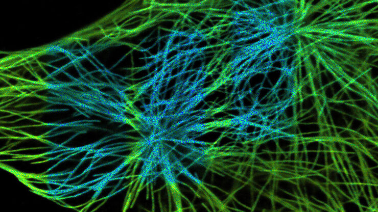
斑马鱼大脑高分辨率全器官成像
结构信息是理解复杂生物系统的关键,而脊椎动物的中枢神经系统是最复杂的生物结构之一。要想从发育中的斑马鱼身上分离出一个完整的大脑,我们需要覆盖大约10平方毫米的区域,深度在毫米范围内。通常,低倍透镜不能提供足够的分辨率来揭示神经组织中复杂结构之间的相互作用。此外,由于散射过程,使用共聚焦显微镜在致密生物组织内成像深度通常限制在大约10微米。

Loading...

从光到思维:共聚焦显微镜中的检测器和测量技术
本文概述了共聚焦显微镜中常用的重要传感器。“共聚焦显微镜”在此特指“真共聚焦扫描”,即仅对单点进行照明和测量的技术。本文旨在为用户提供不同技术之间清晰的概览,并针对不同应用场景给出合适的传感器选择建议,而非深入探讨专业细节。
