
Filter articles
标签
产品
Loading...

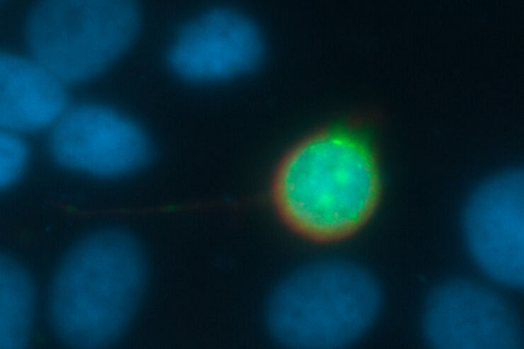
Overcoming Challenges with Microscopy when Imaging Moving Zebrafish Larvae
Zebrafish is a valuable model organism with many beneficial traits. However, imaging a full organism poses challenges as it is not stationary. Here, this case study shows how zebrafish larvae can be…
Loading...

Aneurysm Clipping: Assessing Perforators in Real-time with AR Fluorescence
This article covers two aneurysm clipping cases highlighting the clinical benefits of GLOW800 Augmented Reality Fluorescence application in neurosurgery, based on insights from Prof. Tohru Mizutani,…
Loading...

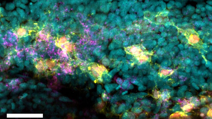
Advancing Uterine Regenerative Therapies with Endometrial Organoids
Prof. Kang's group investigates important factors that determine the uterine microenvironment in which embryo insertion and pregnancy are successfully maintained. They are working to develop new…
Loading...
![[Translate to chinese:] Optical microscope image, which is a composition of both brightfield and fluorescence illumination, showing organic contamination on a wafer surface. The inset images in the upper left corner show the brightfield image (above) and fluorescence image (below with dark background).](/fileadmin/_processed_/a/1/csm_Organic_contamination_on_a_wafer_surface_6699165cee.jpg "[Translate to chinese:] Optical microscope image, which is a composition of both brightfield and fluorescence illumination, showing organic contamination on a wafer surface.")
晶圆上的光刻胶残留和有机污染物的可视化
随着半导体上集成电路(IC)的尺寸低于10纳米,在晶圆检测中有效检测光刻胶残留等有机污染物和缺陷变得越来越重要。光学显微镜仍然是常见的检测方法,但对于有机污染物,明场和其他类型的照明可能会存在局限性。本文讨论了荧光显微镜如何在半导体行业的QC、故障分析和研发过程中有效检测晶圆上的光刻胶残留和其他有机污染物。
Loading...

激光显微切割技术用于组织和细胞分离的协议 - 免费下载电子书
激光显微切割(LMD,也称为激光捕获显微切割或LCM)使用户能够分离特定的单个细胞或整个组织区域,甚至亚细胞结构如染色体。纯化的组织和细胞可用于下游的RNA、DNA和蛋白质组工作流程。
Loading...

数码显微镜摄像头和图像分析的基础技术术语定义
现今绝大多数显微镜都配置了摄像头。摄像头的特征通常决定了所采集到的图像是否能够揭示出研究人员希望观察到的现象。但深入到摄像头术语时,技术术语十分繁杂。我们汇总整理了最为重要的术语及其简明释意以便提供方向。这些术语按字母顺序排列。
Loading...

荧光入门介绍
荧光是George Gabriel Stokes于1852年首次报道的一种现象。他观察到萤石在紫外线照射后开始发光。荧光是光致发光的一种形式,是指一种材料被光照射后会发射出光子。发射光的波长比激发光更长。这种效应又称为斯托克斯位移。
Loading...
超越反卷积
宽场荧光显微镜通常用于视觉呈现生命科学样本中的结构并获取重要信息。利用荧光蛋白或染料,以高度特异性的方式标记离散的样本部分。为了充分了解某种结构,可能需要以三维方式呈现,但这会对使用显微镜带来某些挑战。
Loading...
如何从根本上简化成像设备的工作流程
本集MicaCam中,来自伦敦大学学院(UCL)的特邀嘉宾Christopher Thrasivoulou博士将从成像设备的角度讨论使用Mica的优势。他将讨论如何简化复杂生物系统的成像工作流程并实现自动化。这有助于科学家节省为获取有意义的量化分析结果而投入的时间和精力。为了举例说明此类工作流程,他还会展示如何对荧光标记的固定斑马鱼胚胎进行多色成像。
