Filter articles
标签
产品
Loading...

From Bench to Beam: A Complete Correlative Cryo Light Microscopy Workflow
In the webinar entitled "A Multimodal Vitreous Crusade, a Cryo Correlative Workflow from Bench to Beam" a team of experts discusses the exciting world of correlative workflows for structural biology…
Loading...
![[Translate to chinese:] Block-face created by automatic trimming under fluorescence. Mammalian cells of interest, stained with CellTrackerTM Green are visualized within the block-face using the UC Enuity. [Translate to chinese:] Block-face created by automatic trimming under fluorescence. Mammalian cells of interest, stained with CellTrackerTM Green are visualized within the block-face using the UC Enuity equipped with the stereo microscope M205 FA. In the background a carbon finder grid in black is visible. All samples in the article are created by Felix Gaedke, PhD, CECAD, Cologne, Germany.](/fileadmin/_processed_/c/c/csm_Block_face_created_by_automatic_trimming_using_fluorescence_527c668077.jpg)
如何在块面中自动获取感兴趣的荧光细胞
本文介绍了使用超薄切片超薄切片机自动修整修块功能,获取树脂块面中带有荧光信号的细胞结构。我们展示了如何使用配置有体视显微镜 M205 FA 的超薄切片超薄切片机 UC Enuity ,来识别感兴趣的荧光细胞,如何自动修整包含细胞的块面,以及如何在切片中观察细胞而无需转移到外部显微镜。
Loading...
![UC Enuity [Translate to chinese:] UC Enuity](/fileadmin/_processed_/e/9/csm_UC_Enuity_Detail_Header_411ca2e94e.jpg)
通过自动切片改善您的超薄切片工作流程
在不断发展的电镜样品制备领域,保持领先地位至关重要。这个网络研讨会提供了关于超薄切片最新进展的重要见解,这些进展可以显著增强您实验室的能力。
Loading...
![[Translate to chinese:] Camera image during auto alignment. The feedback lines indicate if the correct edges in the image are detected. Green: Vertical center line [Translate to chinese:] Camera image during auto alignment. The feedback lines indicate if the correct edges in the image are detected. Green: Vertical center line; Magenta: Upper edge of the light gap; White: Lower edge of the light gap (not visible here, falling together with red line); Red: Knife edge; Blue: Left and right edge of the block face being automatically detected.](/fileadmin/_processed_/d/1/csm_Camera_image_during_auto_alignment_UC_Enuity_5e6b7b9057.jpg)
高质量超薄切片:样品与切片刀自动对齐
超薄切片技术是获取样品切片的最常用方法。在室温条件制备时,将样品小块嵌入环氧树脂中,然后通过修剪去除多余的树脂,并使用玻璃刀或金刚石刀将样品切成厚度为50-100纳米之间的薄片。
Loading...
![[Translate to chinese:] Material sample with a large height, size, and weight being observed with an inverted microscope. [Translate to chinese:] Material sample with a large height, size, and weight being observed with an inverted microscope.](/fileadmin/_processed_/f/4/csm_Inverted_microscope_with_large_sample_crop_9ca60cecc7.jpg)
工业应用中倒置显微镜相较于正置显微镜的五大优势
使用倒置显微镜时,您需要从下方观察样本,因为倒置显微镜的光学元件位于样本下方,而使用正置显微镜时,您需要从上方观察样本。一直以来,倒置显微镜主要用于生命科学研究,因为重力将样本沉入含有水性溶液的托座底部,从上方则无法观察到太多内容。但近段时间以来,倒置显微镜在工业应用中也变得越来越流行。我们现在一起来了解倒置显微镜在工业应用中的优势。
Loading...
![[Translate to chinese:] Image of an integrated-circuit (IC) chip cross section acquired at higher magnification showing a region of interest. [Translate to chinese:] Image of an integrated-circuit (IC) chip cross section acquired at higher magnification showing a region of interest.](/fileadmin/_processed_/a/c/csm_IC_chip_cross_section_c413bcc998.jpg)
横截面切片法分析IC芯片的结构与化学成分
从本文中了解如何通过横截面分析法对集成电路 (IC) 芯片等电子元件进行有效的结构和元素分析。探索如何通过研磨系统进行铣削、锯切、磨削和抛光工艺以及用于同时进行目视检测和化学分析的二合一解决方案来完成的。可针对电子行业的各种工作流程和应用实现快速、详细的材料分析,包括竞争分析、质量控制 (QC)、故障分析 (FA) 以及研发 (R&D)。
Loading...

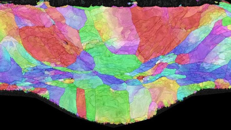
高质量EBSD样品制备
本文介绍了一种使用宽离子束研磨技术为“混合”晶体材料制备可靠且有效的EBSD(电子背散射衍射)样品的方法。该方法产生的横截面具有高质量表面,这对于EBSD分析至关重要。电子背散射衍射(EBSD)材料分析是通过扫描电子显微镜(SEM)进行的。制备混合材料(CPU或铝(Al)、金刚石和石墨(C)的复合材料)的横截面,使其具有适合EBSD分析的高质量表面,可能是一个挑战。


![[Translate to chinese:] Section ribbons with increasing section thickness - silver to purple ending in blue sections. [Translate to chinese:] Section ribbons with increasing section thickness - silver to purple ending in blue sections.](/fileadmin/_processed_/7/2/csm_Section_ribbons_with_increasing_section_thickness_d0e10995bf.jpg)
