
Filter articles
标签
产品
Loading...

Dive into Pancreatic Cancer Research with Big Data
Pancreatic cancer, with a mortality rate near 40%, is challenging to treat due to its proximity to major organs. This story explores the complex biology of pancreatic ductal adenocarcinoma (PDAC),…
Loading...

Uncover the Hidden Complexity of Colon Cancer with Big Data
Colorectal cancer poses a significant health burden. While surgery is effective initially, some patients develop recurrent secondary disease with poor prognosis, necessitating advanced therapies like…
Loading...

Mapping Tumor Immune Landscape with AI-Powered Spatial Proteomics
Spatial mapping of untreated tumors provides an overview of the tumor immune architecture, useful for understanding therapeutic responses. Immunocompetent murine models are essential for identifying…
Loading...

组织中的精密空间蛋白质组学信息
尽管可使用基于成像和质谱的方法进行空间蛋白质组学研究,但是图像与单细胞分辨率蛋白丰度测量值的关联仍然是个巨大的挑战。最近引入的一种方法,深层视觉蛋白质组学(DVP),将细胞表型的人工智能图像分析与自动化的单细胞或单核激光显微切割及超高灵敏度的质谱分析结合在了一起。DVP在保留空间背景的同时,将蛋白丰度与复杂的细胞或亚细胞表型关联在一起。
Loading...
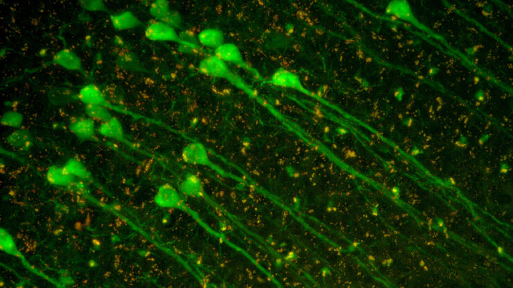
![[Translate to chinese:] Multiplexed Cell DIVE imaging of Adult Human Alzheimer’s brain tissue section demonstrating expression of markers specific to astrocytes (GFAP, S100B), microglia (TMEM119, IBA1), AD-associated markers (p-Tau217, β-amyloid) and immune cells such as CD11b+, CD163+, CD4+, and HLA-DRA+, clustered around the β-amyloid plaques.](/fileadmin/_processed_/b/b/csm_Alzheimers_brain_tissue_section_showing_astrocytes_microglia_immune_cells_6f24036a9f.jpg "[Translate to chinese:] Multiplexed Cell DIVE imaging of Adult Human Alzheimer’s brain tissue section demonstrating expression of markers specific to astrocytes , microglia , AD-associated markers and immune cells clustered around the β-amyloid plaques")
Spatial Analysis of Neuroimmune Interactions in Alzheimer’s Disease
Alzheimer’s disease (AD) is a complex neurodegenerative disorder characterized by neurofibrillary tangles, β-amyloid plaques, and neuroinflammation. These dysfunctions trigger or are exacerbated by…
Loading...

A Guide to Spatial Biology
What is spatial biology, and how can researchers leverage its tools to meet the growing demands of biological questions in the post-omics era? This article provides a brief overview of spatial biology…
Loading...
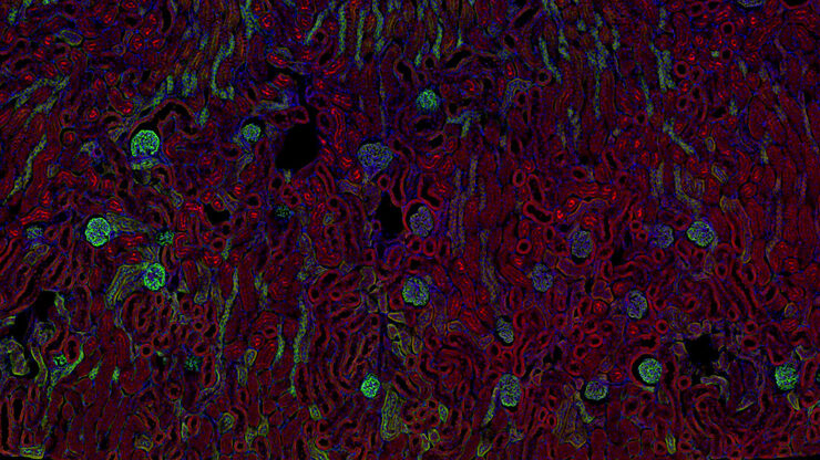
![[Translate to chinese:] Multiplexed Cell DIVE imaging to characterize the spatial landscape in Human Alzheimer’s Cortical Tissue](/fileadmin/_processed_/7/2/csm_Human_Alzheimers_Cortical_Tissue_Multiplexed_Cell_DIVE_imaging_e4cf382069.jpg "[Translate to chinese:] Multiplexed Cell DIVE imaging to characterize the spatial landscape in Human Alzheimer’s Cortical Tissue")
使用空间多重化探测人类阿尔茨海默病皮层切片
阿尔茨海默病(AD)是最常见的神经退行性疾病,其特征是认知功能的逐渐下降。对 AD 大脑的空间分析可能揭示细胞关系,从而促进对疾病病因的更好理解。本研究捕捉了 AD 皮层组织成分的全球概述,并强调了 Cell DIVE 成像的简化工作流程,从数据采集到使用 Aivia 软件的基于人工智能的分析,最终实现更快的洞察。
