
Filter articles
标签
产品
Loading...
A Guide to Super-Resolution
Find out more about Leica super-resolution microscopy solutions and how they can empower you to visualize in fine detail subcellular structures and dynamics.
Loading...
![[Translate to chinese:] Multicolor fixed STED image. Inner ear section, mouse, TauSTED Xtend 589 on AF488 and TauSTED Xtend 775 on AF633-Phalloidin. Sample courtesy of Dennis Derstrof, Klinik für Hals-, Nasen und Ohrenheilkunde, Universität Marburg & Prof. Dr. Dominik Oliver aus dem Institut für Physiologie und Pathophysiologie, Abteilung für Neurophysiologie, Universität Marburg.](/fileadmin/_processed_/c/a/csm_Inner_ear_section_mouse_multicolor_fixed_TauSTED_Xtend_09ebb898fd.jpg "[Translate to chinese:] Multicolor fixed STED image. Inner ear section, mouse, TauSTED Xtend 589 on AF488 and TauSTED Xtend 775 on AF633-Phalloidin.")
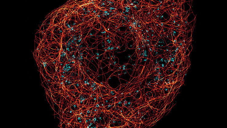
细胞活成像的纳米级扩展
新的STED显微技术方法——TauSTED Xtend,使得在纳米级别下对活体完整样本进行扩展多色成像成为可能。通过结合空间和寿命信息,TauSTED Xtend提供了额外一层信息,允许在极低的光剂量下分辨小细节并在整体结构中解析它们。
Loading...
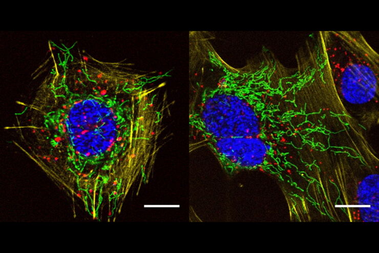
![[Translate to chinese:] Multicolor TauSTED Xtend 775 for Cell Biology applications that require nanoscopy resolution for multiple cellular components. Cells showing vimentin fibrils (AF 594), actin network (ATTO 647N), and nuclear pore basket (CF 680R). Sample courtesy of Brigitte Bergner, Mariano Gonzales Pisfil, Steffen Dietzel, Core Facility Bioimaging, Biomedical Center, Ludwig-Maximilians-University, Munich, Germany.](/fileadmin/_processed_/5/4/csm_Triple_color_fixed_sample_TauSTED_Xtend_2_3_695f805a53.jpg "[Translate to chinese:] Multicolor TauSTED Xtend 775 for Cell Biology applications that require nanoscopy resolution for multiple cellular components. Cells showing vimentin fibrils (AF 594), actin network (ATTO 647N), and nuclear pore basket (CF 680R).")
STED样品制备指南
这份指南旨在帮助用户优化受激发射损耗(STED)纳米成像的样品制备,特别是在使用徕卡微系统的STED显微镜时。它提供了单色STED成像用荧光标记的概述,并对其性能进行了评级。
![[Translate to chinese:] Five-color FLIM-STED](/fileadmin/_processed_/a/3/csm_5-color_FLIM-STED_a783161004.jpg "[Translate to chinese:] Five-color FLIM-STED")
Loading...
A Versatile Palette of Fluorescent Probes
Researchers at the Max Planck Institute for Medical Research in Heidelberg have developed a general strategy to synthesize live-cell compatible fluorogenic probes, and the result are the new MaP (Max…
Loading...

结合 STED 和Lifetime的优势
在这次访谈中,Alberto Diaspro教授讨论了白光激光的优势以及STELLARIS 8 STED的TauSTED技术能力。他分享了自己在使用TauSense、荧光寿命成像和相位分析技术进行科研项目时,与共聚焦系统相关的经验。
Loading...

超分辨率显微镜图片库
由于光的衍射极限,传统共聚焦显微镜无法分辨约240纳米以下的结构。当需要提高分辨率以研究衍射极限尺度以下的结构和分子事件时,会使用超分辨率显微镜技术,如STED、PALM或STORM,或某些解卷积处理方法。
Loading...

Regulators of Actin Cytoskeletal Regulation and Cell Migration in Human NK Cells
Dr. Mace will describe new advances in our understanding of the regulation of human NK cell actin cytoskeletal remodeling in cell migration and immune synapse formation derived from confocal and…
Loading...

显微镜在病毒学中的应用
引起新型冠状病毒肺炎(Covid-19)的冠状病毒SARS-CoV-2肆虐全球并影响了我们生活的方方面面。对于免疫和治疗方法的搜索研究(即如何抗击该病毒)成为了2020年全人类的第一要务。显微镜在这类研究中起着重要作用。为了了解受体结合、基因组释放、复制、装配和病毒出芽的基本原理以及我们的免疫系统效应,可以使用不同的方法和显微镜。本文概述了为什么显微镜是病毒学和感染生物学的重要工具,并举例说明了不…
