Filter articles
标签
产品
Loading...
![[Translate to chinese:] Multiplexed Cell DIVE imaging of Adult Human Alzheimer’s brain tissue section demonstrating expression of markers specific to astrocytes , microglia , AD-associated markers and immune cells clustered around the β-amyloid plaques [Translate to chinese:] Multiplexed Cell DIVE imaging of Adult Human Alzheimer’s brain tissue section demonstrating expression of markers specific to astrocytes (GFAP, S100B), microglia (TMEM119, IBA1), AD-associated markers (p-Tau217, β-amyloid) and immune cells such as CD11b+, CD163+, CD4+, and HLA-DRA+, clustered around the β-amyloid plaques.](/fileadmin/_processed_/b/b/csm_Alzheimers_brain_tissue_section_showing_astrocytes_microglia_immune_cells_6f24036a9f.jpg)
Spatial Analysis of Neuroimmune Interactions in Alzheimer’s Disease
Alzheimer’s disease (AD) is a complex neurodegenerative disorder characterized by neurofibrillary tangles, β-amyloid plaques, and neuroinflammation. These dysfunctions trigger or are exacerbated by…
Loading...

Revealing Neuronal Migration’s Molecular Secrets
Different approaches can be used to investigate neuronal migration to their niche in the developing brain. In this webinar, experts from The University of Oxford present the microscopy tools and…
Loading...
![[Translate to chinese:] Multiplexed Cell DIVE imaging to characterize the spatial landscape in Human Alzheimer’s Cortical Tissue [Translate to chinese:] Multiplexed Cell DIVE imaging to characterize the spatial landscape in Human Alzheimer’s Cortical Tissue](/fileadmin/_processed_/7/2/csm_Human_Alzheimers_Cortical_Tissue_Multiplexed_Cell_DIVE_imaging_e4cf382069.jpg)
使用空间多重化探测人类阿尔茨海默病皮层切片
阿尔茨海默病(AD)是最常见的神经退行性疾病,其特征是认知功能的逐渐下降。对 AD 大脑的空间分析可能揭示细胞关系,从而促进对疾病病因的更好理解。本研究捕捉了 AD 皮层组织成分的全球概述,并强调了 Cell DIVE 成像的简化工作流程,从数据采集到使用 Aivia 软件的基于人工智能的分析,最终实现更快的洞察。
Loading...
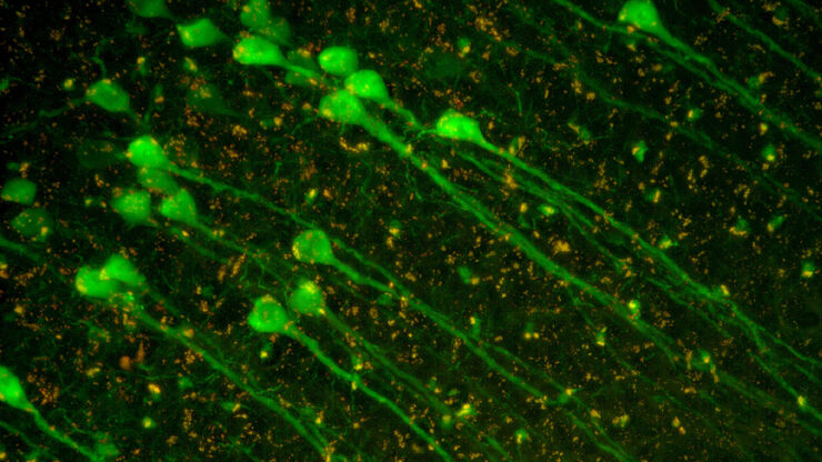
![[Translate to chinese:] Image of murine dopaminergic neurons which have been marked for laser microdissection (LMD). [Translate to chinese:] Image of murine dopaminergic neurons which have been marked for laser microdissection (LMD).](/fileadmin/_processed_/b/f/csm_Murine_dopaminergic_neurons_marked_for_LMD_d784dbd83f.jpg)
利用激光显微切割(LMD)在空间背景下分离神经元
在阿尔茨海默病之后,帕金森病是第二常见的进行性神经退行性疾病。在首发症状出现之前,中脑中高达70%的多巴胺释放神经元已经死亡。本文描述了如何使用现代激光显微切割(LMD)方法帮助解决帕金森病之谜。研究涉及在空间背景下分离和分析神经元。这些细胞来自帕金森病患者的死后黑质组织样本,以便深入了解该病的分子机制。
Loading...

激光显微切割技术如何助力神经科学研究取得开创性进展?
玛尔塔·帕特林尼博士,卡罗林斯卡学院的高级科学家,分享了她在成人人类神经发生开创性研究中使用激光显微切割(LMD)的经验,并提供了关于LMD在空间蛋白质组学和精准医学中未来应用潜力的个人见解。
Loading...

激光显微切割技术用于组织和细胞分离的协议 - 免费下载电子书
激光显微切割(LMD,也称为激光捕获显微切割或LCM)使用户能够分离特定的单个细胞或整个组织区域,甚至亚细胞结构如染色体。纯化的组织和细胞可用于下游的RNA、DNA和蛋白质组工作流程。
Loading...
![[Translate to chinese:] The role of extracellular signalling mechanisms in the correct development of the human brain [Translate to chinese:] The role of extracellular signalling mechanisms in the correct development of the human brain](/fileadmin/_processed_/a/e/csm_The_role_of_extracellular_signalling_mechanisms_in_the_correct_development_of_the_human_brain_6b9e3b80f0.jpg)
在神经发育过程中,细胞是如何相互交流的?
细胞间通信是大脑发育过程中一个必不可少的过程,它受到多种因素的影响,包括细胞的形态、粘附分子、局部细胞外基质和分泌囊泡。在本次网络研讨会上,您将了解到对这些机制更深入的理解是如何推动对神经发育障碍的理解的。
Loading...

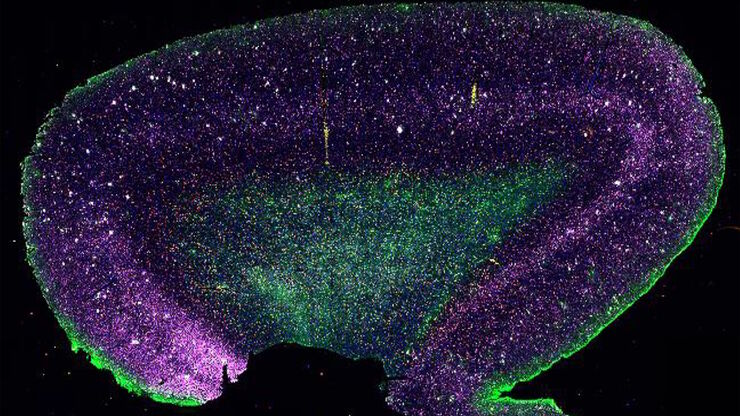
大脑的形状:阿尔茨海默病的空间生物学
阿尔茨海默病(AD)是一种神经退行性疾病,也是导致晚年认知障碍的常见原因。阿尔茨海默病的特征是出现含β-淀粉样蛋白的斑块和含磷酸化 tau 的神经纤维缠结。目前尚缺乏治疗和预防AD的有效疗法。我们将Cell DIVE与Cell Signaling Technology的抗体结合使用,检查了AD的突触过程并从空间上确定了神经胶质细胞和神经元等细胞,证明了超多标免疫荧光成像技术可用于探测AD大脑。


