
Filter articles
标签
产品
Loading...

How does Real-time OCT Imaging Impact Precision in Corneal Surgery?
Corneal surgery is a highly specialized field. It requires great surgical precision to overcome challenges such as visualizing clearly the full anterior chamber, performing Descemet membrane peeling…
Loading...
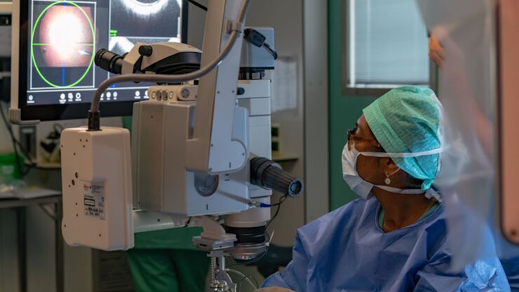
![[Translate to chinese:] How the M822 microscope enhances surgical precision in eye surgeries - Insights from Dr. Dhami. Image courtesy of Dr. Abhinav Dhami.](/fileadmin/_processed_/d/3/csm_Dr_Abhinav_Dhami_with_M822_631e107586.jpg "[Translate to chinese:] How the M822 microscope enhances surgical precision in eye surgeries - Insights from Dr. Dhami. Image courtesy of Dr. Abhinav Dhami.")
听听Dhami 医生关于购买眼科显微镜的专业见解
在本文中,了解来自印度北部的眼科手术顾问Abhinav Dhami医生如何使用Leica Microsystems的M822眼科显微镜来提高他的手术精确度,以及哪些关键特性使他决定购买这款眼科显微镜用于他的实践。
Loading...

Dislocated Cataract Angle Closure Aided by Intraoperative OCT
Learn how a dislocated cataract was treated with angle closure assisted by intraoperative OCT to achieve long-term good results without future lens dislocation.
Loading...

术中OCT引导的青光眼支架修复手术
青光眼是导致全球不可逆失明的主要原因之一。小梁网切除术和导管分流引流术等历史悠久的手术技术会带来巨大的短期风险和潜在并发症。近年来,随着微创青光眼手术(MIGS)的出现,手术方法有了长足的发展,其特点是对组织的破坏最小、内路粘小管植入、手术时间短、器械简单、术后恢复快。
![[Translate to chinese:] Prof. Nikolaos Bechrakis uses the Proveo 8 ceiling mounted microscope with EnFocus intraoperative OCT. Images provided by Prof. Nikolaos Bechrakis.](/fileadmin/_processed_/7/3/csm_Prof_Bechrakis_uses_Proveo_8_ceiling_mounted_microscope_with_EnFocus_intraoperative_OCT_0d79cea5a2.jpg "[Translate to chinese:] Prof. Nikolaos Bechrakis uses the Proveo 8 ceiling mounted microscope with EnFocus intraoperative OCT. Images provided by Prof. Nikolaos Bechrakis.")
![[Translate to chinese:] Keratoplasty of pathologic cornea](/fileadmin/_processed_/6/d/csm_Pathologic_cornea_633aed9eab.jpg "[Translate to chinese:] Keratoplasty of pathologic cornea")
Loading...
![[Translate to chinese:] Dr. Ozana Moraru shares two primary open-angle glaucoma cases in which trabeculectomy bleb needling was performed using the Leica M844 microscope with EnFocus intraoperative OCT. Image courtesy of Dr. Ozana Moraru.](/fileadmin/_processed_/1/0/csm_Trabeculectomy_bleb_needling_performed_with_Leica_M844_microscope_with_EnFocus_intraoperative_OCT_14e8587e0d.jpg "[Translate to chinese:] Dr. Ozana Moraru shares two primary open-angle glaucoma cases in which trabeculectomy bleb needling was performed using the Leica M844 microscope with EnFocus intraoperative OCT. Image courtesy of Dr. Ozana Moraru.")
术中光学显像如何帮助青光眼手术获得更多洞察力
青光眼是导致全球失明的主要原因之一。青光眼手术可以延缓疾病的发展。在青光眼手术过程中,术中光学相干断层扫描(OCT)的使用为眼科外科医生提供了更佳的可视化效果,让他们更深入地了解表面下组织对手术操作的反应。 莫拉鲁博士通过两个原发性开角型青光眼(POAG)的临床病例强调了它的价值。
Loading...

眼科: 复杂白内障手术中的可视化
白内障手术是最常见的眼科手术。为了满足白内障手术的需要,Ozana Moraru 博士使用了 Leica Microsystems 的 M844 显微镜和 EnFocus 术中光学相干断层扫描 (OCT) 以及 3D 可视化系统。在本案例研究中,她介绍了术中光学相干断层扫描如何为标准和复杂的白内障手术病例提供有用信息。
