Filter articles
标签
产品
Loading...
![[Translate to chinese:] 3D-volume-rendered light-sheet microscope image of a spheroid showing depth coding in different colors. [Translate to chinese:] 3D-volume-rendered light-sheet microscope image of a spheroid showing depth coding in different colors.](/fileadmin/_processed_/4/8/csm_Spheroid_showing_depth_coding_in_different_colors_3D_DLS_ff196ec61f.jpg)
利用DLS对细胞球中的抗癌药物摄取进行成像
细胞球3D细胞培养模型模拟了活组织的生理和功能,使其成为研究肿瘤形态和筛选抗癌药物的有用工具。药物AZD2014是一种公认的哺乳动物雷帕霉素靶蛋白(mTOR)通路抑制剂[1]。mTOR的异常激活会促进肿瘤生长和转移,导致AZD2014进入临床试验作为抗癌分子。其具体的抗肿瘤机制尚不清楚。
Loading...

利用多重中频成像设计您的研究课题
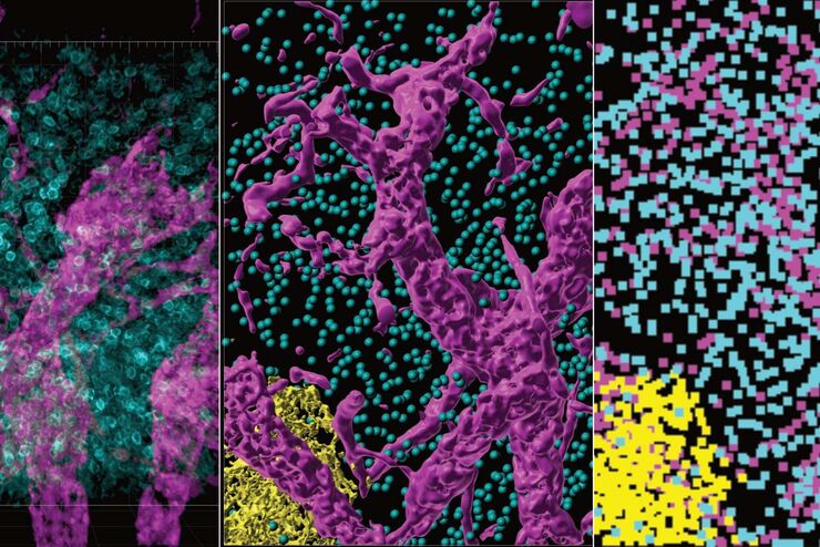
多重组织分析是一种功能强大的技术,可对单个固定组织样本中的细胞类型位置和细胞类型相互作用进行比较。在多重分析研究开始之前,研究人员通常会提出以下问题: "我如何知道组织中哪些生物标记物是相关的?另外,随着研究问题的发展,我如何转向其他生物标记物?巧妙的研究设计有助于回答现有的问题,并能继续探索研究开始时并不明显的新联系。
Loading...

Cell DIVE已验证的抗体将使您对实验结果产生信心
Cell DIVE超多标组织成像分析整体解决方案包括经严格验证的350+抗体资源库,高灵敏度高特异性的应用于Cell DIVE循环染色成像中。抗体验证的方法可以帮助您找到合适的抗体以及最佳的实验条件,快速的让您开展超多标成像分析的实验。抗体库中的每种抗体都经过严格的三步验证过程,(a)评估在FFPE上的表现性能;(b)确定其最佳的染色条件以及是否可用作直标抗体;(c)探究由于Cell…
Loading...

优化 THUNDER 平台以实现高内涵玻片扫描
随着对全组织成像需求的不断增长以及对不同生物标本中 FL 信号定量的需要,HC 成像技术的极限受到了考验,而核心设备的用户可培训性和易用性则成为了成本和效率的问题。在这里,我们展示了在我们的设施中为THUNDER平台开发的可行工作流程,以支持从 KO-小鼠组织分析到人类癌症的各种研究环境需求。
Loading...

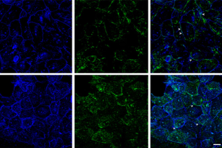
Regulators of Actin Cytoskeletal Regulation and Cell Migration in Human NK Cells
Dr. Mace will describe new advances in our understanding of the regulation of human NK cell actin cytoskeletal remodeling in cell migration and immune synapse formation derived from confocal and…
Loading...

改进成像技术以了解细胞器膜细胞动态
了解正常组织和肿瘤组织中的细胞功能,是推动潜在治疗策略研究和了解某些治疗失败原因的关键因素。单细胞分析在生物医学研究中至关重要,它能揭示在癌症等复杂疾病中哪些细胞和分子通路发生了改变。
Loading...

从器官到组织再到细胞:使用宽场显微镜分析 3D 标本
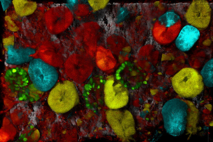
在传统的宽场显微镜下从厚的三维样本中获取高质量的数据和图像是具有挑战性的,因为存在失焦光的干扰。在本次网络研讨会中,Falco Krüger 展示了THUNDER成像仪如何通过Computational Clearing技术使这一切成为可能。
Loading...

Rodent and Small-Animal Surgery
Learn how you can perform rodent (mouse, rat, hamster) and small-animal surgery efficiently with a microscope for developmental biology and medical research applications by reading this article.


