Filter articles
标签
产品
Loading...
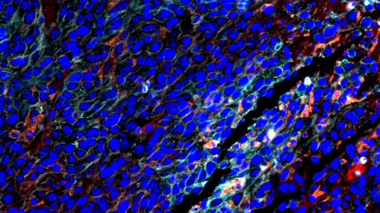
![[Translate to chinese:] Multiplexed Cell DIVE imaging of Colon Adenocarcinoma (CAC) tissue. [Translate to chinese:] Multiplexed Cell DIVE imaging of Colon Adenocarcinoma (CAC) tissue. A panel of approximately 30 biomarkers targeted towards various leukocyte lineages, epithelial, stromal, and endothelial cell types was utilized to characterize the tumor immune microenvironment in human colon adenocarcinoma (CAC) tissue.](/fileadmin/_processed_/c/7/csm_Colon_Adenocarcinoma_CAC_tissue_multiplexed_Cell_DIVE_image_33055bc13b.jpg)
通过成像和AI绘制结直肠癌的景观
结肠癌是一种高负担疾病。尽管进行了化疗干预和手术切除,但疾病可能会复发。了解结肠癌微环境对于改善治疗效果是必要的。在这里,我们使用空间生物学方法,通过Cell DIVE和 Aivia可视化结肠腺癌组织中的30个生物标志物。我们探讨了肿瘤组织的血管化、免疫细胞反应和细胞增殖。
Loading...
![[Translate to chinese:] Esophageal tissue with a squamous cell carcinoma labelled with the 4 biomarkers PanCk, DAPI, NaKATPase, and Vimentin. [Translate to chinese:] Esophageal tissue with a squamous cell carcinoma labelled with the 4 biomarkers PanCk, DAPI, NaKATPase, and Vimentin.](/fileadmin/_processed_/7/1/csm_Esophageal_Squamous_Cell_Carcinoma_4_Markers_7ac3f29e4b.jpg)
探索多重生物成像如何推进癌症研究
观看行业和学术专家进行的内容丰富的讨论,分享他们在研究中使用多重成像技术的知识。了解多重成像技术如何通过发现以前难以捉摸的分子洞察力,彻底改变肿瘤学、神经学和免疫学。利用先进的成像技术深入了解组织微环境,从而对代谢紊乱和癌症等疾病有新的认识。
Loading...

Coherent Raman Scattering Microscopy Publication List
CRS (Coherent Raman Scattering) microscopy is an umbrella term for label-free methods that image biological structures by exploiting the characteristic, intrinsic vibrational contrast of their…
Loading...
![[Translate to chinese:] Branched organoid growing in collagen where the Nuclei are labeled blue. To detect the mechanosignaling process, the YAP1 is labeled green. [Translate to chinese:] Branched organoid growing in collagen where the Nuclei are labeled blue. To detect the mechanosignaling process, the YAP1 is labeled green.](/fileadmin/_processed_/a/e/csm_Branched_organoid_growing_in_collagen_dc289aa8c6.jpg)
检查癌症类器官的发展进程
德国慕尼黑工业大学的Andreas Bausch实验室研究细胞和生物体中不同结构和功能形成的细胞和生物物理机制。他的团队设计了新的策略、方法和分析工具,以量化微米和纳米等级的发展机制和动态过程。关键研究领域包括干细胞和类器官,从乳腺类器官到胰腺癌类器官,以更好地了解疾病模型。


![[Translate to chinese:] Pancreatic Ductal Adenocarcinoma with 11 Aerobic Glycolysis/Warburg Effect biomarkers shown – BCAT, Glut1, HK2, HTR2B, LDHA, NaKATPase, PCAD, PCK26, PKM2, SMA1, and Vimentin. [Translate to chinese:] Pancreatic Ductal Adenocarcinoma with 11 Aerobic Glycolysis/Warburg Effect biomarkers shown – BCAT, Glut1, HK2, HTR2B, LDHA, NaKATPase, PCAD, PCK26, PKM2, SMA1, and Vimentin.](/fileadmin/_processed_/d/d/csm_Pancreatic_Ductal_Adenocarcinoma_11_Aerobic_Glycolysis_Markers_ROI3_074851c0a4.jpg)
![[Translate to chinese:] Spheroid stained with Cyan: Dapi nuclear countertain; Green AF488 Involucrin; Orange AF55 Phalloidin Actin; Magenta AF647 CK14. [Translate to chinese:] Spheroid stained with Cyan: Dapi nuclear countertain; Green AF488 Involucrin; Orange AF55 Phalloidin Actin; Magenta AF647 CK14.](/fileadmin/_processed_/6/8/csm_Spheroid_stained_with_Cyan_4color_overlay_78dff87b83.jpg)
![[Translate to chinese:] Hepatocellular Carcinoma with 13 biomarkers shown – Beta-Catenin, CD3D, CD4, CD8a, CD31, CD44, CD163, DAPI, PanCK, PCK26, PD1, SMA, and Vimentin. [Translate to chinese:] Hepatocellular Carcinoma with 13 biomarkers shown – Beta-Catenin, CD3D, CD4, CD8a, CD31, CD44, CD163, DAPI, PanCK, PCK26, PD1, SMA, and Vimentin.](/fileadmin/_processed_/5/b/csm_Hepatocellular_Carcinoma_13_Markers_Zoom2_f5674cfd25.jpg)

