
Filter articles
标签
产品
Loading...
Capture life as it happens
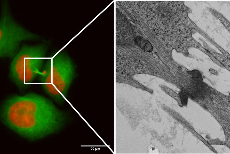
With the Leica Nano Workflow, searching for the needle in the haystack is a thing of the past. Take advantage of correlative light and electron microscopy to identify directly the right cell at the…
Loading...
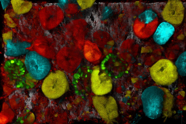
![[Translate to chinese:] C. elegans Gonades - THUNDER Imager Adult hermaphrodit, Staining: blue - DAPI (Nucleus), green - SP56 (sperms), red - RME-2 (oocyte), mangenta - PGL-1 (RNA + protein granules) Image courtesy of Prof. Dr. Christian Eckmann, Martin Luther University, Halle, Germany](/fileadmin/_processed_/3/c/csm_THUNDER-Imager_C-elegans_Gonades_Physiology_3600_59ce4afec3.jpg "[Translate to chinese:] C. elegans Gonades")
生理学图片库
生理学是关于生物体内的过程和功能。生理学研究的重点是生物体器官、组织或细胞的活动和功能,包括所涉及的物理和化学现象。在此,我们以不同的样本为例,向您展示与生理学有关的图片。
Loading...
![[Translate to chinese:] Nematostella](/fileadmin/_processed_/d/c/csm_Nematostella-LiveImaging-Stellaris_19b3bde47a.jpg "[Translate to chinese:] Nematostella")
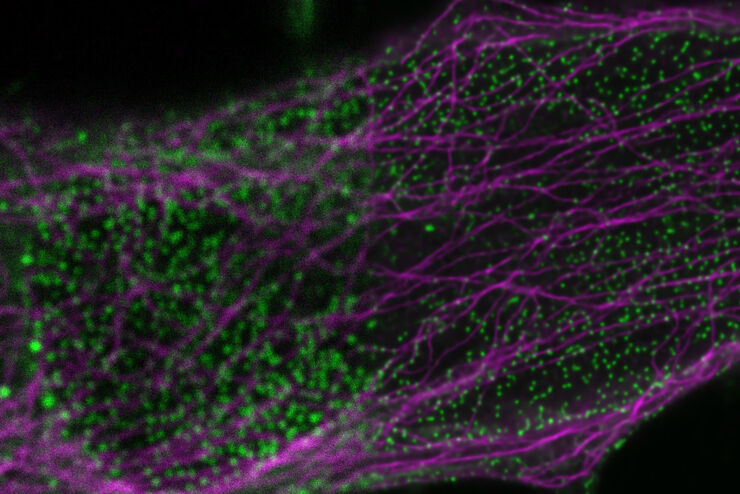
活细胞成像图库
活细胞显微镜技术是更好地了解细胞和分子功能的基础。如今,宽场显微镜是用于长时间观察细胞动态和发育的最常用技术。共聚焦显微镜也是一种重要工具,可生成三维结构图像,并以高空间和时间分辨率研究高度动态的细胞过程,同时使标本保持接近原生状态。
Loading...

将动态活细胞数据融入超微结构
采用徕卡Nano的工作流程,可以避免过去如海底捞针似的寻找。利用光电关联显微技术,在适当的时间直接鉴别出正确的细胞,并将动态的活细胞数据融入其超微结构中。
 and astrocytes (green) in a cortical spheroid derived from human induced pluripotent stem cells.")
Loading...
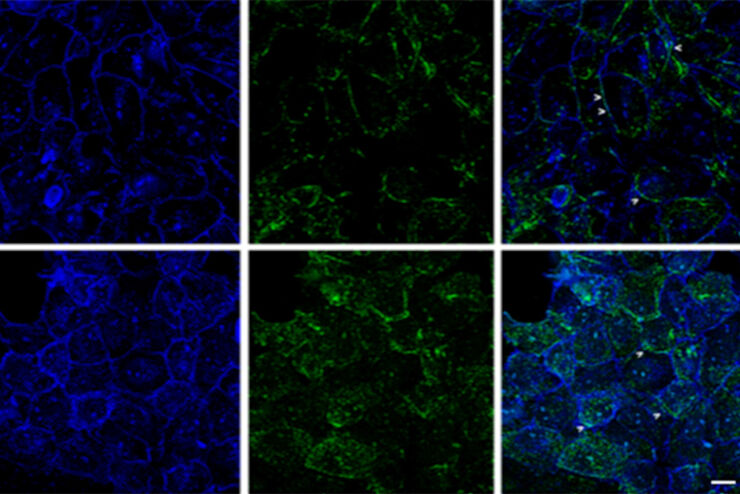
改进成像技术以了解细胞器膜细胞动态
了解正常组织和肿瘤组织中的细胞功能,是推动潜在治疗策略研究和了解某些治疗失败原因的关键因素。单细胞分析在生物医学研究中至关重要,它能揭示在癌症等复杂疾病中哪些细胞和分子通路发生了改变。
Loading...

Image Gallery: THUNDER Imager
To help you answer important scientific questions, THUNDER Imagers eliminate the out-of-focus blur that clouds the view of thick samples when using camera-based fluorescence microscopes. They achieve…
