Filter articles
标签
产品
Loading...

How to Prepare Samples for Stimulated Raman Scattering (SRS) imaging
Find here guidelines for how to prepare samples for stimulated Raman scattering (SRS), acquire images, analyze data, and develop suitable workflows. SRS spectroscopic imaging is also known as SRS…
Loading...

Coherent Raman Scattering Microscopy Publication List
CRS (Coherent Raman Scattering) microscopy is an umbrella term for label-free methods that image biological structures by exploiting the characteristic, intrinsic vibrational contrast of their…
Loading...

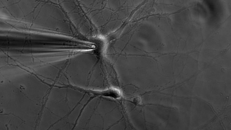
什么是膜片钳技术?
离子通道的生理学一直是神经科学家感兴趣的一个重要话题。诞生于1970年代的膜片钳技术开启了电生理学家的新时代。它不仅可以对整个细胞进行高分辨率电流记录,还可以对切下的细胞膜片进行高分辨率电流记录。甚至可以研究单通道事件。然而,由于需要复杂且高灵敏的设备,广泛的生物学背景和高水平的实验技能,电生理学仍然是最具挑战性的实验室方法之一。
Loading...

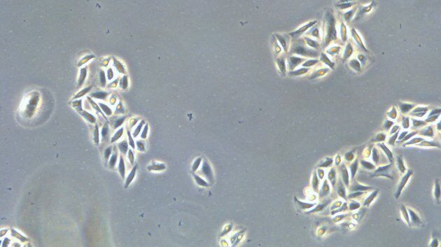
如何使用数字化显微镜测定细胞汇合度
本文介绍了如何以一致性的方式测量细胞汇合度。对于生命科学研究领域,例如癌症生物学、干细胞或再生医学,研究通常需要特定生长条件下的细胞。这些条件包括定期检查的细胞形态和汇合度。
Loading...

如何对细胞培养进行快速正确的检测
本文介绍了对培养贴壁细胞系进行传代的一般工作流程及步骤说明。哺乳类细胞体外培养是在癌症、药物开发、组织工程、干细胞、疾病细胞和分子生物学等生物医学研究领域进行临床和药物研究的重要模型。要想成功维持细胞系,需要通过控制生长条件来维持细胞生理和表型的稳定性。定期监测细胞生长,对细胞进行传代培养以确保连续性。
Loading...

Virtual Reality Showcase for STELLARIS Confocal Microscopy Platform
In this webinar, you will discover how to perform 10-color acquisition using a confocal microscope. The challenges of imaged-based approaches to identify skin immune cells. A new pipeline to assess…

![[Translate to chinese:] THUNDER image of brain-capillary endothelial-like cells derived from human iPSCs (induced pluripotent stem cells) where cyan indicates nuclei and magenta tight junctions. [Translate to chinese:] THUNDER image of brain-capillary endothelial-like cells derived from human iPSCs (induced pluripotent stem cells) where cyan indicates nuclei and magenta tight junctions.](/fileadmin/_processed_/8/e/csm_Brain-capillary_endothelial-like_cells_derived_from_human_iPSCs_dc0fae4c20.jpg)
![[Translate to chinese:] Spheroid stained with Cyan: Dapi nuclear countertain; Green AF488 Involucrin; Orange AF55 Phalloidin Actin; Magenta AF647 CK14. [Translate to chinese:] Spheroid stained with Cyan: Dapi nuclear countertain; Green AF488 Involucrin; Orange AF55 Phalloidin Actin; Magenta AF647 CK14.](/fileadmin/_processed_/6/8/csm_Spheroid_stained_with_Cyan_4color_overlay_78dff87b83.jpg)
![[Translate to chinese:] Mouse cortical neurons. Transgenic GFP (green). Image courtesy of Prof. Hui Guo, School of Life Sciences, Central South University, China [Translate to chinese:] Mouse cortical neurons. Transgenic GFP (green). Image courtesy of Prof. Hui Guo, School of Life Sciences, Central South University, China](/fileadmin/_processed_/2/a/csm_THUNDER_Imager_Mouse_cortical_neuron_1fa1718d8f.jpg)
