
Filter articles
标签
产品
Loading...

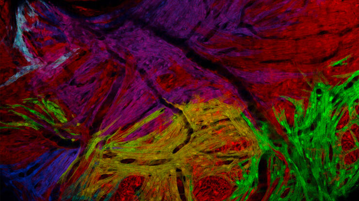
Overcoming Challenges with Microscopy when Imaging Moving Zebrafish Larvae
Zebrafish is a valuable model organism with many beneficial traits. However, imaging a full organism poses challenges as it is not stationary. Here, this case study shows how zebrafish larvae can be…
Loading...
斑马鱼研究
为了在筛选、分拣、操作和成像过程中获取高质量结果,您需要观察细节和结构,从而为您的下一步研究做出正确的决策。
徕卡体视显微镜和透射光底座以出众的光学器件和优良的分辨率而闻名,是全世界研究学者的首选。
Loading...
如何从根本上简化成像设备的工作流程
本集MicaCam中,来自伦敦大学学院(UCL)的特邀嘉宾Christopher Thrasivoulou博士将从成像设备的角度讨论使用Mica的优势。他将讨论如何简化复杂生物系统的成像工作流程并实现自动化。这有助于科学家节省为获取有意义的量化分析结果而投入的时间和精力。为了举例说明此类工作流程,他还会展示如何对荧光标记的固定斑马鱼胚胎进行多色成像。
![[Translate to chinese:] Zebrafish heart showing the ventricle with an injury in the lower area](/fileadmin/_processed_/9/6/csm_Zebrafish_heart_showing_ventricle_with_injury_teaser_6b564c5217.jpg "[Translate to chinese:] Zebrafish heart showing the ventricle with an injury in the lower area")
Loading...
![[Translate to chinese:] Light sheet microscopy tilescan of zebrafish.](/fileadmin/_processed_/a/d/csm_DLS_Sample_Preparation_zebrafish_tilescan_intro_d8ebec802d.jpg "[Translate to chinese:] Light sheet microscopy tilescan of zebrafish.")
使用 U 形玻璃毛细管进行样品装载
徕卡显微系统的DLS显微镜系统是一种创新概念,将光片显微技术集成到共聚焦平台中。由于其独特的光学结构,样本可以安装在标准玻璃底培养皿上,与传统的安装程序相比,几乎不需要或只需很少的适应。在这里,我们介绍了一种便捷的方法,能够快速准备样本以进行光片成像。
Loading...

Imaging and Analyzing Zebrafish, Medaka, and Xenopus
Discover how to image and analyze zebrafish, medaka, and Xenopus frog model organisms efficiently with a microscope for developmental biology applications from this article.
