
Filter articles
标签
产品
Loading...
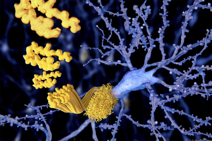
阿尔茨海默斑块:厚切片中的快速可视化
超过 60%的所有诊断为痴呆症的病例归因于阿尔茨海默病。该疾病的典型特征是脑组织的组织学改变。目前尚无治愈该疾病的方法。一些治疗方法试图减缓致命的进程或缓解患者的症状。梅赫达德·沙姆鲁博士的实验室研究病理性脑功能,旨在为阿尔茨海默病的新疗法的发现做出贡献。他们使用这种疾病的小鼠模型研究炎症在阿尔茨海默病进展中的作用。这需要对厚的未清除脑组织进行成像。
Loading...
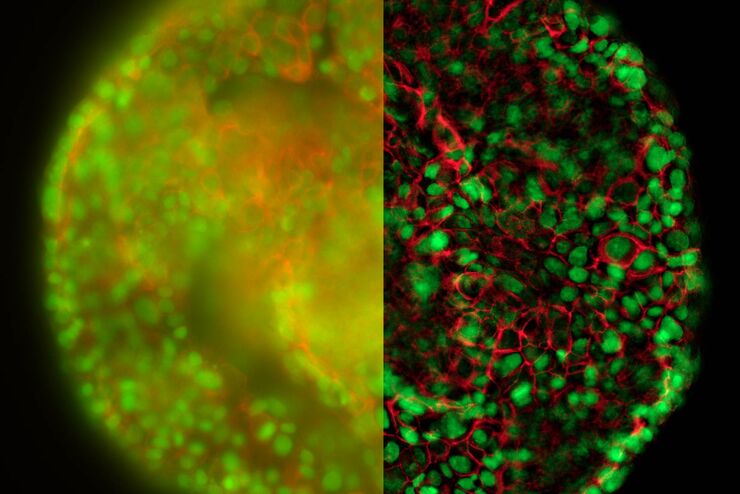
清晰对比、无雾的 3D 样本实时图像
历史上,宽场显微镜并不适合对大样本/标本体积进行成像。图像背景(BG)主要来源于观察样本的失焦区域,显著降低了成像系统的对比度、有效动态范围和最大可能的信噪比(SNR)。记录的图像显示出典型的雾霭,并且在许多情况下,无法提供进一步分析所需的细节水平。处理厚三维样本的研究人员要么使用替代显微镜方法,要么尝试通过后处理一系列图像来减少雾霭。
![[Translate to chinese:]](/fileadmin/_processed_/5/7/csm_convalaria_Widefield-topic_Intro_7c19cc9054.jpg "[Translate to chinese:]")
Loading...
![[Translate to chinese:] HeLa cells stimulated with LPS. Image has been subjected to deconvolution.](/fileadmin/_processed_/c/0/csm_HeLa_cells_stimulated_with_LPS_deconvolution_cf0b3cf706.jpg "[Translate to chinese:] HeLa cells stimulated with LPS. Image has been subjected to deconvolution.")
显微镜下的慢性炎症
在慢性炎症的过程中,身体的某些部位会反复发炎。许多人类疾病都是如此。在宽场光学显微镜的帮助下,可以对从细胞水平到整个生物体的潜在过程进行检查。本文介绍了几种宽场显微镜应用,如免疫荧光、活细胞成像、组织学和比率分析,以深入了解慢性炎症的发展、相关疾病及其治疗。
Loading...

无限远光学系统
“无限远光学”这一概念是指在显微镜的物镜和镜筒透镜之间具有平行光线的光束路径。平面光学元件可以进入到这个“无限远空间”中,而不影响成像,这对于利用DIC或荧光等对比度方法至关重要。
现代显微技术需要在无限远光路中添加多种光学仪器,如光源或激光装置。满足这一需求的不同方法已经出现,本文对其进行了描述。
Loading...
![[Translate to chinese:] Eukaryotic cells](/fileadmin/_processed_/3/1/csm_Widefield_Application_Letter_Fura_01_Sep07_en-1_8f3a569916.jpg "[Translate to chinese:] Eukaryotic cells")
使用钙指示剂 Fura2 的宽场钙成像
在真核细胞中,Ca2+是信号转导通路中最广泛使用的第二信使之一。细胞内的 Ca2+ 水平通常保持较低,因为 Ca2+ 常常与磷酸化和羧酸化化合物形成不溶性复合物。通常,细胞质中的 Ca2+ 浓度在 100 nM 的范围内。作为对刺激的反应,Ca2+ 可以从外部介质或内部储存释放,以提高 Ca2+ 浓度。
Loading...
![[Translate to chinese:] QTM B, 1963, the first commercial automated image analysis system for microscope images, based on a TV camera and developed by Metals Research in Cambridge, England.](/fileadmin/_processed_/8/a/csm_QTM_B_1963_cut_08146176de.jpg "[Translate to chinese:] QTM B, 1963, the first commercial automated image analysis system for microscope images, based on a TV camera and developed by Metals Research in Cambridge, England.")
图像分析 50 年
现代图像分析系统对来自自动显微镜和数码相机的图像执行高度复杂的图像处理功能。50 年前,第一套图像分析系统是模拟系统,以摄像机为基础,面积测量可通过仪表读取。不过,它标志着这一领域自动化的开端。
Loading...
Image Processing for Widefield Microscopy
Fluorescence microscopy is a modern and steadily evolving tool to bring light to current cell biological questions. With the help of fluorescent proteins or dyes it is possible to make discrete…
Loading...

Applications of TIRF Microscopy in Life Science Research
The special feature of TIRF microscopy is the employment of an evanescent field for fluorophore excitation. Unlike standard widefield fluorescence illumination procedures with arc lamps, LEDs or…
