
Filter articles
标签
产品
Loading...
![[Translate to chinese:] Mouse lung sections](/fileadmin/_processed_/c/3/csm_Studying_Pulmonary_Fibrosis_Teaser_4d06e4f265.jpg)
肺纤维化研究
本文中所示结果表明,相比明场,使用偏振光可以更清晰地分辨小鼠肺组织中的胶原蛋白纤维化和非纤维化区域。为更好地理解促使瘢痕组织形成的肺纤维化,正常情况下会研究组织中的纤维化区域。分别使用明场和偏振光对小鼠肺组织中的胶原蛋白纤维化病变区域进行成像。成像胶原蛋白时,使用一般的染色法和明场显微镜很难区分纤维化和非纤维化区域。本文中使用偏振光对肺组织进行成像,两个区域呈现出非常明显的颜色差异。
Loading...

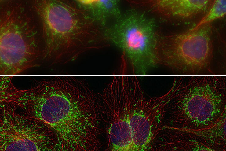
Image Gallery: THUNDER Imager
To help you answer important scientific questions, THUNDER Imagers eliminate the out-of-focus blur that clouds the view of thick samples when using camera-based fluorescence microscopes. They achieve…
Loading...
从器官到组织再到细胞:使用宽场显微镜分析 3D 标本
在传统的宽场显微镜下从厚的三维样本中获取高质量的数据和图像是具有挑战性的,因为存在失焦光的干扰。在本次网络研讨会中,Falco Krüger 展示了THUNDER成像仪如何通过Computational Clearing技术使这一切成为可能。
Loading...

An Introduction to Computational Clearing
Many software packages include background subtraction algorithms to enhance the contrast of features in the image by reducing background noise. The most common methods used to remove background noise…
Loading...
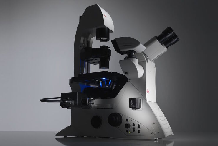
Factors to Consider When Selecting a Research Microscope
An optical microscope is often one of the central devices in a life-science research lab. It can be used for various applications which shed light on many scientific questions. Thereby the…
![[Translate to chinese:] Influenca in lung epithelial cells (porcine) - THUNDER Imager 3D Cell Culture Influenca virus – red, cilia – green, Nuclei – blue.](/fileadmin/_processed_/1/5/csm_THUNDER-Imager-3D-Cell-culture_Rabies_brain_ferret_scaled_6db61a7a35.jpg "[Translate to chinese:] Influenca in lung epithelial cells (porcine) - THUNDER Imager 3D Cell Culture Influenca virus – red, cilia – green, Nuclei – blue.")
Loading...

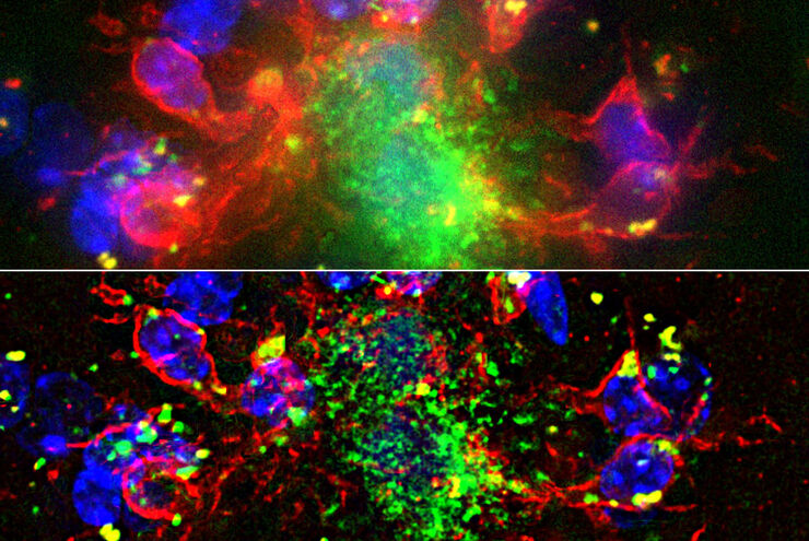
显微镜在病毒学中的应用
引起新型冠状病毒肺炎(Covid-19)的冠状病毒SARS-CoV-2肆虐全球并影响了我们生活的方方面面。对于免疫和治疗方法的搜索研究(即如何抗击该病毒)成为了2020年全人类的第一要务。显微镜在这类研究中起着重要作用。为了了解受体结合、基因组释放、复制、装配和病毒出芽的基本原理以及我们的免疫系统效应,可以使用不同的方法和显微镜。本文概述了为什么显微镜是病毒学和感染生物学的重要工具,并举例说明了不…
Loading...
Computational Clearing - 增强3D标本成像
本次网络研讨会旨在阐明有助于THUNDER显微成像仪实现三维样品可视化的关键规格,并改进研究人员的成像相关工作流程。
Loading...
THUNDER成像:高效、灵活、易操作,让您的日常成像工作流更轻松
本次网络研讨会将展示 THUNDER 在许多不同生命科学应用中的多功能性和性能:从计数视网膜切片中的细胞核和癌组织切片中的 RNA 分子,到监测阿拉伯芥幼苗中的钙波等等。
