Filter articles
标签
产品
Loading...
![[Translate to chinese:] Identification of distinct structures_roundworm_Ascaris_female [Translate to chinese:] Identification of distinct structures_roundworm_Ascaris_female](/fileadmin/_processed_/a/b/csm_Identification_of_distinct_structures_roundworm_Ascaris_female_eddbe9bbff.jpg)
从概览中查找相关样本细节
在从图像到图像的搜索中切换到快速查看整个样本概览,并即刻识别重要的样本细节。利用这些知识,使用载玻片、培养皿和多孔板的模板自动设置高分辨率图像采集。LAS X Navigator软件像是样本细胞的GPS,总能为用户指明通向高质量数据的清晰路径,这是生命科学平台STELLARIS和THUNDER成像仪上的一款强大的导航工具。LAS X Navigator支持将宽场、立体或共聚焦实验与舞台应用相结合。
Loading...
![[Translate to chinese:] Formation of 3D spheroids from 1000 MDCK cells [Translate to chinese:] Formation of 3D spheroids; Time lapse acquisition over 72 hours](/fileadmin/_processed_/7/7/csm_Formation_of_3D_spheroids_from_MX1-GFP_cells_66a26e09c9.jpg)
高效的长期延时拍摄技术
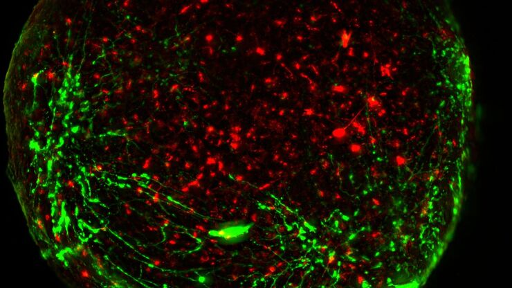
当对球状体做延时拍摄技术时,会出现某些挑战。由于实验可能持续数天,必须实现长时间的样本存活,这就需要确保接近生理条件。本文描述的长期延时研究使用了全场景显微成像分析平台MICA来研究U343和MDCK细胞球形成。细胞球生长需要最佳条件,以确保细胞周期和增殖不受干扰。
Loading...

在显微成像和图像分析中运用人工智能和机器学习技术
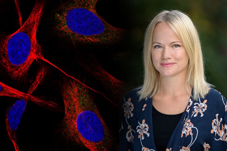
Emma Lundberg 教授是瑞典 KTH 皇家理工学院细胞生物学蛋白质组学教授。她还是细胞图谱项目的总监,该项目是瑞典人类蛋白质图谱(HPA)项目不可或缺的组成部分,后者是用于研究人类蛋白质组的开源资源。细胞图谱项目是 HPA 的一部分,提供人类细胞系中 RNA 和蛋白质的表达及时空分布的高分辨率图像。Lundberg…
Loading...

多通道活细胞成像注意事项
同时多色成像,确保实验成功:活细胞成像实验是了解动态过程的关键。这类实验使我们能够观察记录活体状态下的细胞,而不会可能因固定或终止不同活体过程而产生干扰性伪影。
Loading...
![[Translate to chinese:] HeLa Kyoto cells [Translate to chinese:] HeLa Kyoto cells (HKF1, H2B-mCherry, alpha Tubulin, mEGFP). Left image: Maximum projection of a z-stack prior to ICC and LVCC. Right image: Maximum projection of a mosaic z-stack after ICC and LVCC.](/fileadmin/_processed_/6/a/csm_How_to_improve_live_cell_imaging_with_Leica_Nano_Workflow_teaser_6d36e3e6d8.jpg)
如何使用Coral Life(活细胞光电联用)改进活细胞成像
对于活细胞 CLEM 应用而言,光学显微镜成像是在正确的时间以正确的状态识别正确细胞的关键步骤。在本文中,徕卡专家就使用宽场系统的优势以及使用蓝宝石作为细胞培养基底时需要克服的障碍分享了他们的见解。
Loading...

Dissecting Proteomic Heterogeneity of the Tumor Microenvironment
This lecture will highlight cutting edge applications in applying laser microdissection and microscaled quantitative proteomics and phosphoproteomics to uncover exquisite intra- and inter-tumor…
Loading...
![[Translate to chinese:] Image: Mouse kidney section with Alexa Fluor™ 488 WGA, Alexa Fluor™ 568 Phalloidin, and DAPI. Sample is a FluoCells™ prepared slide #3 from Thermo Fisher Scientific, Waltham, MA, USA. [Translate to chinese:] Mouse kidney section with Alexa Fluor™ 488 WGA, Alexa Fluor™ 568 Phalloidin, and DAPI. Sample is a FluoCells™ prepared slide #3 from Thermo Fisher Scientific, Waltham, MA, USA. Images courtesy of Dr. Reyna Martinez – De Luna, Upstate Medical University, Department of Ophthalmology.](/fileadmin/_processed_/3/a/csm_The_Power_of_Pairing_Adaptive_Deconvolution_teaser_6ab6b726a1.jpg)
自适应反卷积与 Computational Clearing 结合的力量
反卷积是一种计算方法,用于恢复被点扩散函数(PSF)和噪声源破坏的物体图像。在本技术简介中,您将了解徕卡显微系统提供的反卷积算法如何帮助您克服宽视场 (WF) 荧光显微镜中由于光的波动性和光学元件对光的衍射而造成的图像分辨率和对比度损失。探索由用户控制或自动反卷积的方法,查看并解析更多的结构细节。
Loading...

改进成像技术以了解细胞器膜细胞动态
了解正常组织和肿瘤组织中的细胞功能,是推动潜在治疗策略研究和了解某些治疗失败原因的关键因素。单细胞分析在生物医学研究中至关重要,它能揭示在癌症等复杂疾病中哪些细胞和分子通路发生了改变。


