
Filter articles
标签
产品
Loading...
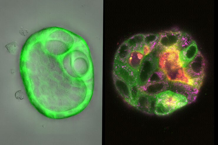
, the cis-golgi matrix protein GM130 (AF488, green), and the trans-golgi network membrane protein TGN46 (AF647, red).")
高尔基组织对细胞应激的反应变化
在本集MicaCam直播活动中,来自海德堡欧洲分子生物学实验室的特邀嘉宾George Galea将对用各类化疗药物进行治疗的HeLa Kyoto细胞进行分析,并观察其对高尔基复合体和细胞核的组织和定位的影响。
Loading...
![[Translate to chinese:] Pancreatic ductal adenocarcinoma tissue section imaged with Cell DIVE](/fileadmin/_processed_/f/8/csm_Pancreatic_ductal_adenocarcinoma_tissue_section_teaser_3e2f21e476.jpg "[Translate to chinese:] Pancreatic ductal adenocarcinoma tissue section imaged with Cell DIVE")
多重成像的类型、优势和应用
与传统显微镜相比,多重成像技术能观察到更多的生物标记物,是一种新兴的、令人兴奋的从人体组织样本中提取信息的方法。通过同时观察多种生物标记物,可以协同探索以前只能单独探索的生物通路,并识别和探测复杂的组织和细胞表型。目前已有许多不同的多重成像方法,每种方法都采用不同的方法来实现更高的复杂性。
Loading...
![[Translate to chinese:] 3D Reconstruction of brain slide image_Mica](/fileadmin/_processed_/b/b/csm_Brain_slide_image_3D_Reconstruction_Mica_teaser_dde26fb597.jpg "[Translate to chinese:] 3D Reconstruction of brain slide image_Mica")
3D组织成像:快速预览到高分辨率成像的一键切换
3D组织成像广泛应用于生命科学领域。研究人员利用它来揭示组织组成和完整性的详细信息,或从实验操作中得出结论,或比较健康与不健康的样本。本文介绍了MICA如何帮助研究人员进行3D组织成像。
Loading...

如何实现快速、稳定的多色活细胞成像
研究人员在活细胞成像技术的帮助下,可以深入了解活细胞甚至完整生物体的动态过程,这包括细胞内和细胞外活动。一些代表性的示例包括蛋白质或脂质转运、免疫细胞迁移,类器官的细胞组织等。活细胞成像要求样本和显微镜系统具备特定的属性。在这篇文章中,我们描述了MICA对这些先决条件的具体调整,并提供了合适的示例。
![[Translate to chinese:] Zebrafish heart showing the ventricle with an injury in the lower area](/fileadmin/_processed_/9/6/csm_Zebrafish_heart_showing_ventricle_with_injury_teaser_6b564c5217.jpg "[Translate to chinese:] Zebrafish heart showing the ventricle with an injury in the lower area")
![[Translate to chinese:] Developing zebrafish (Danio rerio) embryo, from sphere stage to somite stages.](/fileadmin/_processed_/8/f/csm_Developing_Zebrafish_embryo_MicaCam_teaser_81cef69cf1.jpg "[Translate to chinese:] Developing zebrafish (Danio rerio) embryo, from sphere stage to somite stages.")
Loading...

用MICA完成Caspase 3/7多色检测
Caspases与细胞凋亡过程相关,因此可以利用caspase检测来确定细胞是否正在经历这种程序化的细胞死亡。这些检测可以通过例如流式细胞仪、平板读数仪实现,也可以在显微镜上完成,显微镜可为量化数据补充可见的结构信息。在这篇文章中,我们描述了MICA是如何用于caspase 3/7测定。借助Navigator或像素分类器等工具,MICA让设置、执行和分析caspase…
Loading...

如何获得具有完全时空相关性的多标记实验数据
首期MicaCam会聚焦于活细胞实验当中的挑战。我们的主持人Lynne Turnbull和Oliver Schlicker将以活细胞内线粒体活动研究为例,手把手为您展示如何用多孔板培养箱设计您的实验,以及如何分析结果。
