
Filter articles
标签
产品
Loading...
![[Translate to chinese:] Microscopy for neuroscience research](/fileadmin/_processed_/9/5/csm_Microscopy_for_neuroscience_research_6a48c90764.jpg "[Translate to chinese:] Microscopy for neuroscience research")
神经科学显微镜面临哪些挑战?
显微镜是神经科学研究领域的强大工具。不过,当涉及到对神经过程进行成像以及使用不同的样品类型(例如厚神经组织或脑类器官)时,科研人员可能会面临到很多挑战。这本30页的电子书包含众多真实的案例,以讨论我们最常见到的一些挑战,同时展示了如何使用THUNDER 成像技术克服这些挑战。
Loading...
超越反卷积
宽场荧光显微镜通常用于视觉呈现生命科学样本中的结构并获取重要信息。利用荧光蛋白或染料,以高度特异性的方式标记离散的样本部分。为了充分了解某种结构,可能需要以三维方式呈现,但这会对使用显微镜带来某些挑战。
Loading...

精确分析宽视野荧光图像
利用荧光显微镜的特异性,即便是使用厚样品和大尺寸样品,研究人员也能够快速轻松地准确观察和分析生物学过程和结构。然而,离焦荧光会提高背景荧光,降低对比度,影响图像的精确分割。THUNDER 与Aivia 的组合可以有效解决这一问题。前者可以消除图像模糊,后者会使用人工智能技术自动分析宽视野图像,提高操作速度和精确性。下面,我们来详细了解下这一协作方法。
Loading...

High-resolution 3D Imaging to Investigate Tissue Ageing
Award-winning researcher Dr. Anjali Kusumbe demonstrates age-related changes in vascular microenvironments through single-cell resolution 3D imaging of young and aged organs.
Loading...

优化 THUNDER 平台以实现高内涵玻片扫描
随着对全组织成像需求的不断增长以及对不同生物标本中 FL 信号定量的需要,HC 成像技术的极限受到了考验,而核心设备的用户可培训性和易用性则成为了成本和效率的问题。在这里,我们展示了在我们的设施中为THUNDER平台开发的可行工作流程,以支持从 KO-小鼠组织分析到人类癌症的各种研究环境需求。
Loading...
![[Translate to chinese:] C. elegans Gonades - THUNDER Imager Adult hermaphrodit, Staining: blue - DAPI (Nucleus), green - SP56 (sperms), red - RME-2 (oocyte), mangenta - PGL-1 (RNA + protein granules) Image courtesy of Prof. Dr. Christian Eckmann, Martin Luther University, Halle, Germany](/fileadmin/_processed_/3/c/csm_THUNDER-Imager_C-elegans_Gonades_Physiology_3600_59ce4afec3.jpg "[Translate to chinese:] C. elegans Gonades")
生理学图片库
生理学是关于生物体内的过程和功能。生理学研究的重点是生物体器官、组织或细胞的活动和功能,包括所涉及的物理和化学现象。在此,我们以不同的样本为例,向您展示与生理学有关的图片。
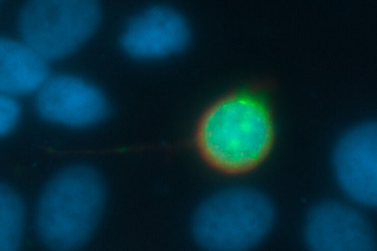
![[Translate to chinese:] Virally labeled neurons (red) and astrocytes (green) in a cortical spheroid derived from human induced pluripotent stem cells. THUNDER Model Organism Imagerwith a 2x 0.15 NA objective at 3.4x zoomwas used to produce this 425 μm Z-stack (26 positions), which is presented here as an Extended Depth of Field(EDoF)projection.](/fileadmin/_processed_/e/c/csm_THUNDER_Imager_Model-Org_Header-Gallery-Neuroscience_e911743fb7.jpg "[Translate to chinese:] Virally labeled neurons (red) and astrocytes (green) in a cortical spheroid derived from human induced pluripotent stem cells.")
![[Translate to chinese:] Pollen Flower - Taken with a 20x/0.8 objective, area of 6mm² with a depth of 100μm. 15 stitched tiles with 4 colors (DAPI/GFP/TRITC/Cy5) - a total of 13020 images. Video courtesy of James Marr, Leica Microsystems, USA](/fileadmin/_processed_/d/1/csm_THUNDER_Imager_Pollen-Flowe_1febb91d34.jpg "[Translate to chinese:] Pollen Flower")
Loading...
![[Translate to chinese:] Cryo FIB lamella - Overlay of SEM and confocal fluorescence image. Target structure in yeast cells (nuclear pore proteine Nup159-Atg8-split Venus, red) marked by an arrow. Scale bar: 5 µm. Alegretti et al., Nature 586, 796-800 (2020).](/fileadmin/_processed_/c/d/csm_Targeting_Nuclear_Pore_Complexes_teaser_27b8f39513.jpg "[Translate to chinese:] Cryo FIB lamella - Overlay of SEM and confocal fluorescence image")
使用冷冻共聚焦显微镜定位活性循环核孔复合物
本文介绍了如何利用冷冻光学显微镜,尤其是冷冻共焦显微镜来提高冷冻工作流程的可靠性。评估了EM网格和样品的质量,并分析了目标结构的分布。本文展示了如何将冷冻共焦3D数据投射到SEM图像上,将感兴趣结构可靠地保留在FIB切割的薄片内,以便在冷冻TEM中进行进一步研究。
