
Filter articles
标签
产品
Loading...
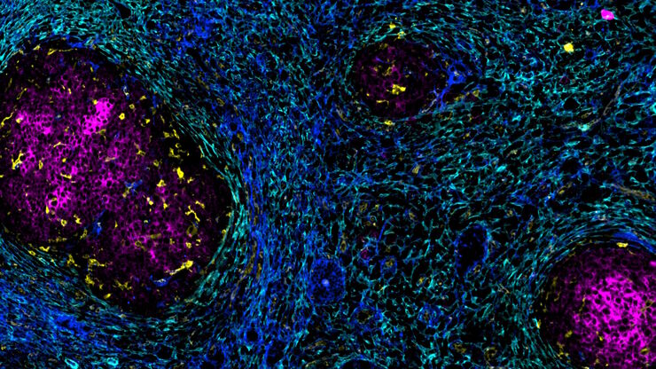
![[Translate to chinese:] Multiplexed Cell DIVE imaging of Colon Adenocarcinoma (CAC) tissue. A panel of approximately 30 biomarkers targeted towards various leukocyte lineages, epithelial, stromal, and endothelial cell types was utilized to characterize the tumor immune microenvironment in human colon adenocarcinoma (CAC) tissue.](/fileadmin/_processed_/c/7/csm_Colon_Adenocarcinoma_CAC_tissue_multiplexed_Cell_DIVE_image_33055bc13b.jpg "[Translate to chinese:] Multiplexed Cell DIVE imaging of Colon Adenocarcinoma (CAC) tissue.")
通过成像和AI绘制结直肠癌的景观
结肠癌是一种高负担疾病。尽管进行了化疗干预和手术切除,但疾病可能会复发。了解结肠癌微环境对于改善治疗效果是必要的。在这里,我们使用空间生物学方法,通过Cell DIVE和 Aivia可视化结肠腺癌组织中的30个生物标志物。我们探讨了肿瘤组织的血管化、免疫细胞反应和细胞增殖。

![[Translate to chinese:] Pancreatic Ductal Adenocarcinoma with 11 Aerobic Glycolysis/Warburg Effect biomarkers shown – BCAT, Glut1, HK2, HTR2B, LDHA, NaKATPase, PCAD, PCK26, PKM2, SMA1, and Vimentin.](/fileadmin/_processed_/d/d/csm_Pancreatic_Ductal_Adenocarcinoma_11_Aerobic_Glycolysis_Markers_ROI3_074851c0a4.jpg "[Translate to chinese:] Pancreatic Ductal Adenocarcinoma with 11 Aerobic Glycolysis/Warburg Effect biomarkers shown – BCAT, Glut1, HK2, HTR2B, LDHA, NaKATPase, PCAD, PCK26, PKM2, SMA1, and Vimentin.")
Loading...
加速不同组织多重成像的发现
组织的多重成像对于肿瘤-免疫相互作用的研究以及人类细胞图谱等发现工作越来越重要。 欢迎加入我们的演讲,Andrea J. Radtke 博士解释了如何使用迭代漂白扩展多重性 (IBEX) 绘制组织图谱,并讨论了用于多重成像的广泛社区资源。
Loading...

Transforming Multiplexed 2D Data into Spatial Insights Guided by AI
Aivia 13 handles large 2D images and enables researchers to obtain deep insights into microenvironment surrounding their phenotypes with millions of detected objects and automatic clustering up to 30…
![[Translate to chinese:] Hepatocellular Carcinoma with 13 biomarkers shown – Beta-Catenin, CD3D, CD4, CD8a, CD31, CD44, CD163, DAPI, PanCK, PCK26, PD1, SMA, and Vimentin.](/fileadmin/_processed_/5/b/csm_Hepatocellular_Carcinoma_13_Markers_Zoom2_f5674cfd25.jpg "[Translate to chinese:] Hepatocellular Carcinoma with 13 biomarkers shown – Beta-Catenin, CD3D, CD4, CD8a, CD31, CD44, CD163, DAPI, PanCK, PCK26, PD1, SMA, and Vimentin.")
Loading...

大脑的形状:阿尔茨海默病的空间生物学
阿尔茨海默病(AD)是一种神经退行性疾病,也是导致晚年认知障碍的常见原因。阿尔茨海默病的特征是出现含β-淀粉样蛋白的斑块和含磷酸化 tau 的神经纤维缠结。目前尚缺乏治疗和预防AD的有效疗法。我们将Cell DIVE与Cell Signaling Technology的抗体结合使用,检查了AD的突触过程并从空间上确定了神经胶质细胞和神经元等细胞,证明了超多标免疫荧光成像技术可用于探测AD大脑。
Loading...
![[Translate to chinese:] Co-detection of 10 extracellular matrix proteins and 3 topographical tissue landmarks by multiplex immunostaining within a single high-grade fibrous hotspot from a human hepatocellular carcinoma](/fileadmin/_processed_/0/8/csm_Single_high-grade_fibrous_hotspot_from_a_human_hepatocellular_carcinoma_ec79be2bf1.jpg "[Translate to chinese:] Co-detection of 10 extracellular matrix proteins and 3 topographical tissue landmarks by multiplex immunostaining within a single high-grade fibrous hotspot from a human hepatocellular carcinoma")
肝细胞癌中癌症干细胞位点的原位鉴定
在这里,我们探索了一种突破性的多重免疫检测方法,通过多重成像对细胞外基质(ECM)特征进行原位定位,从而识别肝细胞癌(HCC)内的癌症干细胞龛。
Loading...
![[Translate to chinese:] Esophageal tissue with a squamous cell carcinoma labelled with the 4 biomarkers PanCk, DAPI, NaKATPase, and Vimentin.](/fileadmin/_processed_/7/1/csm_Esophageal_Squamous_Cell_Carcinoma_4_Markers_7ac3f29e4b.jpg "[Translate to chinese:] Esophageal tissue with a squamous cell carcinoma labelled with the 4 biomarkers PanCk, DAPI, NaKATPase, and Vimentin.")
探索多重生物成像如何推进癌症研究
观看行业和学术专家进行的内容丰富的讨论,分享他们在研究中使用多重成像技术的知识。了解多重成像技术如何通过发现以前难以捉摸的分子洞察力,彻底改变肿瘤学、神经学和免疫学。利用先进的成像技术深入了解组织微环境,从而对代谢紊乱和癌症等疾病有新的认识。
