
Filter articles
标签
产品
Loading...

Transforming Multiplexed 2D Data into Spatial Insights Guided by AI
Aivia 13 handles large 2D images and enables researchers to obtain deep insights into microenvironment surrounding their phenotypes with millions of detected objects and automatic clustering up to 30…
Loading...

人工智能显微图像分析-介绍
人工智能引领的显微图像分析和可视化是用于数据驱动型科学发现的一项强大工具。人工智能技术可以帮助研究人员应对具有挑战性的成像应用,让他们能够从图像中获取更多的信息。
Loading...
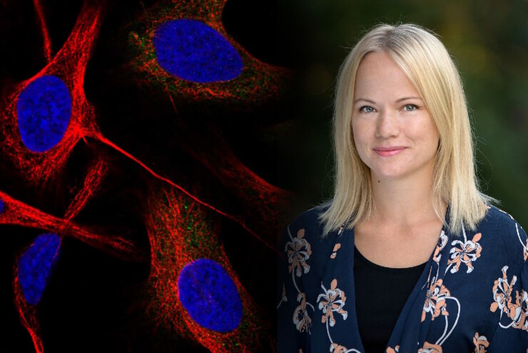
在显微成像和图像分析中运用人工智能和机器学习技术
Emma Lundberg 教授是瑞典 KTH 皇家理工学院细胞生物学蛋白质组学教授。她还是细胞图谱项目的总监,该项目是瑞典人类蛋白质图谱(HPA)项目不可或缺的组成部分,后者是用于研究人类蛋白质组的开源资源。细胞图谱项目是 HPA 的一部分,提供人类细胞系中 RNA 和蛋白质的表达及时空分布的高分辨率图像。Lundberg…
 enhanced with Aivia’s Pixel Classifier (right)")

Loading...
![[Translate to chinese:] C. elegans Gonades - THUNDER Imager Adult hermaphrodit, Staining: blue - DAPI (Nucleus), green - SP56 (sperms), red - RME-2 (oocyte), mangenta - PGL-1 (RNA + protein granules) Image courtesy of Prof. Dr. Christian Eckmann, Martin Luther University, Halle, Germany](/fileadmin/_processed_/3/c/csm_THUNDER-Imager_C-elegans_Gonades_Physiology_3600_59ce4afec3.jpg "[Translate to chinese:] C. elegans Gonades")
生理学图片库
生理学是关于生物体内的过程和功能。生理学研究的重点是生物体器官、组织或细胞的活动和功能,包括所涉及的物理和化学现象。在此,我们以不同的样本为例,向您展示与生理学有关的图片。
Loading...

超分辨率显微镜图片库
由于光的衍射极限,传统共聚焦显微镜无法分辨约240纳米以下的结构。当需要提高分辨率以研究衍射极限尺度以下的结构和分子事件时,会使用超分辨率显微镜技术,如STED、PALM或STORM,或某些解卷积处理方法。
Loading...
组织图片库
对动物和人体组织进行视觉分析对于了解癌症或神经变性等复杂疾病至关重要。从基本的免疫组化到体内成像,共聚焦显微镜和先进的模式可以让人们了解细胞、生物分子及其在环境中的相互作用。
Loading...
![[Translate to chinese:] Nematostella](/fileadmin/_processed_/d/c/csm_Nematostella-LiveImaging-Stellaris_19b3bde47a.jpg "[Translate to chinese:] Nematostella")
活细胞成像图库
活细胞显微镜技术是更好地了解细胞和分子功能的基础。如今,宽场显微镜是用于长时间观察细胞动态和发育的最常用技术。共聚焦显微镜也是一种重要工具,可生成三维结构图像,并以高空间和时间分辨率研究高度动态的细胞过程,同时使标本保持接近原生状态。
