Filter articles
标签
产品
Loading...
![[Translate to chinese:] Fluorescence microscopy image of liver tissue where DNA in the nuclei are stained with Feulgen-pararosanilin and visualized with transmitted green light. [Translate to chinese:] Fluorescence microscopy image of liver tissue where DNA in the nuclei are stained with Feulgen-pararosanilin and visualized with transmitted green light.](/fileadmin/_processed_/0/6/csm_Fluorescence_microscopy_image_of_liver_tissue_0748f2a4d5.jpg)
落射荧光显微镜和反射对比显微镜
多年来,荧光显微镜一直仅使用透射光和暗场照明。随着时间的推移,对改进照明的需求不断增长,这导致了落射照明(也称为入射光照明)的发展。经过 40 年的发展和改进,落射照明荧光显微镜已成为生命科学、临床医学诊断和材料科学领域常规实验室工作和研究的实用方法。大部分开发工作由 Ploem 集团和 Leitz 公司(现为 Leica Microsystems)完成。
Loading...
![[Translate to chinese:] Left: Tissue cells marked with an immunolabel (FITC) illuminated with wide-band UV excitation. Note the tissue structure with blue autofluorescence. Right: Same tissue and same immunostaining with FITC label illuminated with epi-il [Translate to chinese:] Left: Tissue cells marked with an immunolabel (FITC) illuminated with wide-band UV excitation. Note the tissue structure with blue autofluorescence. Right: Same tissue and same immunostaining with FITC label illuminated with epi-illumination using narrow-band blue (490 nm) light. Note the increased image contrast (Ploem, 1967)](/fileadmin/_processed_/c/2/csm_Ploem_Figure_5_Autofluorescence_a_b_fbca553e26.png)
Milestones in Incident Light Fluorescence Microscopy
Since the middle of the last century, fluorescence microscopy developed into a bio scientific tool with one of the biggest impacts on our understanding of life. Watching cells and proteins with the…
Loading...

光学显微镜简史
显微镜的历史始于中世纪。早在11世纪,阿拉伯世界就使用抛光绿柱石制成的平凸透镜作为阅读石来放大手稿。然而,将这些透镜发展成显微镜并非某一个人的功劳,而是众多科学家和学者共同努力的结果。
Loading...
![[Translate to chinese:] QTM B, 1963, the first commercial automated image analysis system for microscope images, based on a TV camera and developed by Metals Research in Cambridge, England. [Translate to chinese:] QTM B, 1963, the first commercial automated image analysis system for microscope images, based on a TV camera and developed by Metals Research in Cambridge, England.](/fileadmin/_processed_/8/a/csm_QTM_B_1963_cut_08146176de.jpg)
图像分析 50 年
现代图像分析系统对来自自动显微镜和数码相机的图像执行高度复杂的图像处理功能。50 年前,第一套图像分析系统是模拟系统,以摄像机为基础,面积测量可通过仪表读取。不过,它标志着这一领域自动化的开端。
Loading...
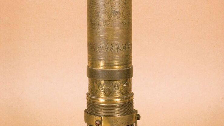
![[Translate to chinese:] A portion of an early binocular microscope developed by John Leonhard Riddel in the early 1850s. [Translate to chinese:] A portion of an early binocular microscope developed by John Leonhard Riddel in the early 1850s.](/fileadmin/_processed_/1/8/csm_Early_binocular_microscope_by_John_Leonhard_Riddel_teaser_9d1ca49ad7.jpg)
体视显微镜的历史
本文概述了从 1600 年至今体视显微镜的发展和演变。直到 19 世纪中叶,所有光学显微镜都是手工制作的。由于无法准确预测透镜的特性,因此必须通过反复试验来制作和测试透镜,直到达到理想的效果。

![[Translate to chinese:] Jellyfish Aequorea Victoria [Translate to chinese:] Jellyfish Aequorea Victoria](/fileadmin/_processed_/7/8/csm_Aequorea3_03_da26f9f6b2.jpg)
