
Filter articles
标签
产品
Loading...

An Introduction to Computational Clearing
Many software packages include background subtraction algorithms to enhance the contrast of features in the image by reducing background noise. The most common methods used to remove background noise…
Loading...
![[Translate to chinese:] Extended Depth of Focus (EDOF) images](/fileadmin/_processed_/5/8/csm_How_to_create_EDOF_images_intro_d6c28929e2.jpg "[Translate to chinese:] Extended Depth of Focus (EDOF) images")
如何创建EDOF(扩展景深)图像
观看此视频,了解如何使用徕卡显微系统LAS X软件的可选扩展景深(EDOF)功能,快速记录具有较大高度变化样本的清晰光学显微镜图像。以使用徕卡显微镜从低倍到高倍拍摄的电路板EDOF图像为例进行了展示。
Loading...
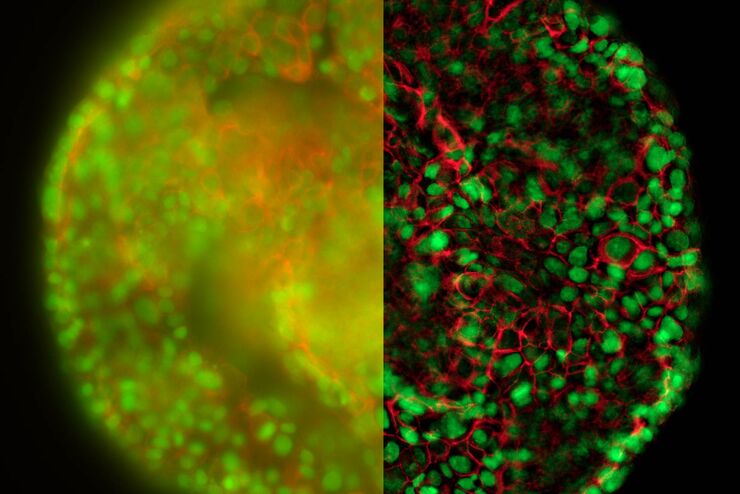
清晰对比、无雾的 3D 样本实时图像
历史上,宽场显微镜并不适合对大样本/标本体积进行成像。图像背景(BG)主要来源于观察样本的失焦区域,显著降低了成像系统的对比度、有效动态范围和最大可能的信噪比(SNR)。记录的图像显示出典型的雾霭,并且在许多情况下,无法提供进一步分析所需的细节水平。处理厚三维样本的研究人员要么使用替代显微镜方法,要么尝试通过后处理一系列图像来减少雾霭。
Loading...
 stereo microscope for a task like surgery.")
Rodent and Small-Animal Surgery
Learn how you can perform rodent (mouse, rat, hamster) and small-animal surgery efficiently with a microscope for developmental biology and medical research applications by reading this article.
Loading...

Imaging and Analyzing Zebrafish, Medaka, and Xenopus
Discover how to image and analyze zebrafish, medaka, and Xenopus frog model organisms efficiently with a microscope for developmental biology applications from this article.
Loading...
![[Translate to chinese:] Fluorescence stereo microscope image of anesthetized Mediterranean fruit flies recorded with a M205 stereo microscope.](/fileadmin/_processed_/1/1/csm_Mediterranean_fruit_flies_recorded_with_M205_FA_1465b7af49.jpg "[Translate to chinese:] Fluorescence stereo microscope image of anesthetized Mediterranean fruit flies recorded with a M205 stereo microscope.")
研究果蝇(黑腹果蝇Drosophila melanogaster)
由于每个实验室的需求可能会有很大的差异,本文展示了科学家和技术人员研究果蝇并使用不同显微镜设置的的实例。此外,基于不同果蝇实验室的经验介绍了推荐的工作流程。本文可以作为建立或扩展果蝇实验室时的参考或指南。
Loading...
![[Translate to chinese:] C. elegans](/fileadmin/_processed_/6/4/csm_C_elegans_6192d4aba6.jpg "[Translate to chinese:] C. elegans")
研究秀丽隐杆线虫(C. elegans)
对于在研究实验室或教室中使用秀丽隐杆线虫(C. elegans)的科学家、技术人员和教师,本报告旨在提供有用的信息,以帮助改进他们的日常工作。其目的是使拾取虫体、转基因、RNA干扰、筛选和功能成像等工作步骤更加高效。本报告还详细介绍了配置研究虫实验室或生物教室/教学实验室的各种可能性,并解释了有关研究虫体方法的内容。
Loading...
![[Translate to chinese:] Forensics microscopy](/fileadmin/_processed_/0/c/csm_Forensics_Microscopy_ec6c3a0561.jpg "[Translate to chinese:] Forensics microscopy")
一条线索都不能少——没有显微镜,就没有法医学
没有线索,就没有犯罪。线索可能很明显,例如犯罪现场的弹壳或者门锁被撬坏的明显痕迹。然而有的时候,线索可能小到微观世界。除了经典的指纹信息,犯罪者还会留下毛发或纤维痕迹。
Loading...
![[Translate to chinese:] QTM B, 1963, the first commercial automated image analysis system for microscope images, based on a TV camera and developed by Metals Research in Cambridge, England.](/fileadmin/_processed_/8/a/csm_QTM_B_1963_cut_08146176de.jpg "[Translate to chinese:] QTM B, 1963, the first commercial automated image analysis system for microscope images, based on a TV camera and developed by Metals Research in Cambridge, England.")
图像分析 50 年
现代图像分析系统对来自自动显微镜和数码相机的图像执行高度复杂的图像处理功能。50 年前,第一套图像分析系统是模拟系统,以摄像机为基础,面积测量可通过仪表读取。不过,它标志着这一领域自动化的开端。
