
Filter articles
标签
产品
Loading...

精确分析宽视野荧光图像
利用荧光显微镜的特异性,即便是使用厚样品和大尺寸样品,研究人员也能够快速轻松地准确观察和分析生物学过程和结构。然而,离焦荧光会提高背景荧光,降低对比度,影响图像的精确分割。THUNDER 与Aivia 的组合可以有效解决这一问题。前者可以消除图像模糊,后者会使用人工智能技术自动分析宽视野图像,提高操作速度和精确性。下面,我们来详细了解下这一协作方法。
Loading...

High-resolution 3D Imaging to Investigate Tissue Ageing
Award-winning researcher Dr. Anjali Kusumbe demonstrates age-related changes in vascular microenvironments through single-cell resolution 3D imaging of young and aged organs.
Loading...

优化 THUNDER 平台以实现高内涵玻片扫描
随着对全组织成像需求的不断增长以及对不同生物标本中 FL 信号定量的需要,HC 成像技术的极限受到了考验,而核心设备的用户可培训性和易用性则成为了成本和效率的问题。在这里,我们展示了在我们的设施中为THUNDER平台开发的可行工作流程,以支持从 KO-小鼠组织分析到人类癌症的各种研究环境需求。
Loading...
![[Translate to chinese:] C. elegans Gonades - THUNDER Imager Adult hermaphrodit, Staining: blue - DAPI (Nucleus), green - SP56 (sperms), red - RME-2 (oocyte), mangenta - PGL-1 (RNA + protein granules) Image courtesy of Prof. Dr. Christian Eckmann, Martin Luther University, Halle, Germany](/fileadmin/_processed_/3/c/csm_THUNDER-Imager_C-elegans_Gonades_Physiology_3600_59ce4afec3.jpg "[Translate to chinese:] C. elegans Gonades")
生理学图片库
生理学是关于生物体内的过程和功能。生理学研究的重点是生物体器官、组织或细胞的活动和功能,包括所涉及的物理和化学现象。在此,我们以不同的样本为例,向您展示与生理学有关的图片。
![[Translate to chinese:] Pollen Flower - Taken with a 20x/0.8 objective, area of 6mm² with a depth of 100μm. 15 stitched tiles with 4 colors (DAPI/GFP/TRITC/Cy5) - a total of 13020 images. Video courtesy of James Marr, Leica Microsystems, USA](/fileadmin/_processed_/d/1/csm_THUNDER_Imager_Pollen-Flowe_1febb91d34.jpg "[Translate to chinese:] Pollen Flower")
Loading...
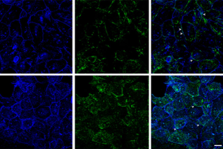
![[Translate to chinese:] Mouse kidney section with Alexa Fluor™ 488 WGA, Alexa Fluor™ 568 Phalloidin, and DAPI. Sample is a FluoCells™ prepared slide #3 from Thermo Fisher Scientific, Waltham, MA, USA. Images courtesy of Dr. Reyna Martinez – De Luna, Upstate Medical University, Department of Ophthalmology.](/fileadmin/_processed_/3/a/csm_The_Power_of_Pairing_Adaptive_Deconvolution_teaser_6ab6b726a1.jpg "[Translate to chinese:] Image: Mouse kidney section with Alexa Fluor™ 488 WGA, Alexa Fluor™ 568 Phalloidin, and DAPI. Sample is a FluoCells™ prepared slide #3 from Thermo Fisher Scientific, Waltham, MA, USA.")
自适应反卷积与 Computational Clearing 结合的力量
反卷积是一种计算方法,用于恢复被点扩散函数(PSF)和噪声源破坏的物体图像。在本技术简介中,您将了解徕卡显微系统提供的反卷积算法如何帮助您克服宽视场 (WF) 荧光显微镜中由于光的波动性和光学元件对光的衍射而造成的图像分辨率和对比度损失。探索由用户控制或自动反卷积的方法,查看并解析更多的结构细节。
Loading...
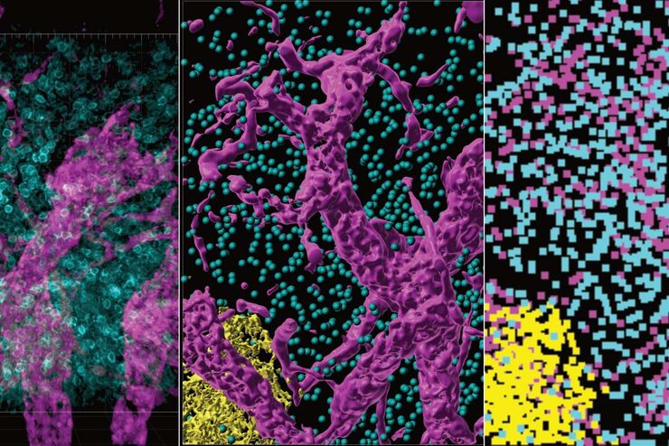
改进成像技术以了解细胞器膜细胞动态
了解正常组织和肿瘤组织中的细胞功能,是推动潜在治疗策略研究和了解某些治疗失败原因的关键因素。单细胞分析在生物医学研究中至关重要,它能揭示在癌症等复杂疾病中哪些细胞和分子通路发生了改变。
Loading...

Image Gallery: THUNDER Imager
To help you answer important scientific questions, THUNDER Imagers eliminate the out-of-focus blur that clouds the view of thick samples when using camera-based fluorescence microscopes. They achieve…
Loading...
从器官到组织再到细胞:使用宽场显微镜分析 3D 标本
在传统的宽场显微镜下从厚的三维样本中获取高质量的数据和图像是具有挑战性的,因为存在失焦光的干扰。在本次网络研讨会中,Falco Krüger 展示了THUNDER成像仪如何通过Computational Clearing技术使这一切成为可能。
