Filter articles
标签
产品
Loading...
![[Translate to chinese:] GLP-1 and PYY localized to distinct secretory pools in L-cells. [Translate to chinese:] GLP-1 and PYY localized to distinct secretory pools in L-cells.](/fileadmin/_processed_/0/2/csm_L-cells_366ce08e69.jpg)
前沿成像技术用于 GPCR 信号传导
通过这个按需网络研讨会,提升您的药理研究,了解 GPCR 信号传导,并探索旨在理解 GPCR 信号如何转化为细胞和生理反应的尖端成像技术。发现领先的研究,扩展我们对这些关键通路的认识,以寻找新的药物发现途径。
Loading...

共聚焦多色成像在癌症研究和免疫学中的潜力
在本次网络研讨会上,来自莫纳什制药科学研究所的CameronNowell和他的同事将分享他们在多重成像方面的经验,以及他们通过巧妙的共聚焦成像采集和利用FLIM等其他多重成像模式所取得的成果。
Loading...
![[Translate to chinese:] Lifetime-based multiplexing in live cells using TauSeparation. Mammalian cells expressing LifeAct-GFP (ibidi GmbH) and labelled with MitoTracker Green. [Translate to chinese:] Lifetime-based multiplexing in live cells using TauSeparation. Mammalian cells expressing LifeAct-GFP (ibidi GmbH) and labelled with MitoTracker Green. Acquisition with one detector, intensity information shown in grey. The two markers can be separated using lifetime information: LifeAct-GFP (cyan), MitoTracker Green (magenta). Image acquired with STELLARIS 5.](/fileadmin/_processed_/8/7/csm_Lifetime-based_multiplexing_in_live_cells_using_TauSeparation_c5d58adf13.jpg)
可重复性、协作和新成像技术的力量
在本次网络研讨会上,您将了解到影响显微镜可重复性的因素,有哪些资源和举措可用于改善显微镜教育并提高其严谨性和可重复性以及研究人员、成像科学家和显微镜供应商之间的合作如何推动创新和采用新技术。
Loading...

人工智能显微成像能够高效检测稀有事件
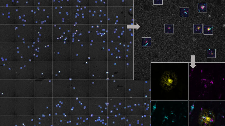
对稀有事件进行定位和选择性成像是许多生物样本研究过程的关键。然而,由于时间限制和高度的复杂性,有些实验无法做到,从而限制了获得新发现的前景。通过基于人工智能的显微成像检测稀有事件,这种工作流程将智能样本导航、图像采集工具和人工智能驱动的图像分析等不同功能融合起来共同协作,能够克服上述局限性。
Loading...

Virtual Reality Showcase for STELLARIS Confocal Microscopy Platform
In this webinar, you will discover how to perform 10-color acquisition using a confocal microscope. The challenges of imaged-based approaches to identify skin immune cells. A new pipeline to assess…

![[Translate to chinese:] Spheroid stained with Cyan: Dapi nuclear countertain; Green AF488 Involucrin; Orange AF55 Phalloidin Actin; Magenta AF647 CK14. [Translate to chinese:] Spheroid stained with Cyan: Dapi nuclear countertain; Green AF488 Involucrin; Orange AF55 Phalloidin Actin; Magenta AF647 CK14.](/fileadmin/_processed_/6/8/csm_Spheroid_stained_with_Cyan_4color_overlay_78dff87b83.jpg)
![[Translate to chinese:] In vivo imaging of a mouse pial and cortical vasculature through a glass window (ROSAmT/mG::Pdgfb-CreERT2 mouse meningeal and cortical visualization following tamoxifen induction and craniotomy). Courtesy: Thomas Mathivet, PhD [Translate to chinese:] In vivo imaging of a mouse pial and cortical vasculature through a glass window (ROSAmT/mG::Pdgfb-CreERT2 mouse meningeal and cortical visualization following tamoxifen induction and craniotomy). Courtesy: Thomas Mathivet, PhD](/fileadmin/_processed_/6/3/csm_Mouse_pial_and_cortical_vasculature_d3e7a9948c.jpg)
![[Translate to chinese:] Five-color FLIM-STED [Translate to chinese:] Five-color FLIM-STED](/fileadmin/_processed_/a/3/csm_5-color_FLIM-STED_a783161004.jpg)

