
Filter articles
标签
产品
Loading...

From Bench to Beam: A Complete Correlative Cryo Light Microscopy Workflow
In the webinar entitled "A Multimodal Vitreous Crusade, a Cryo Correlative Workflow from Bench to Beam" a team of experts discusses the exciting world of correlative workflows for structural biology…
Loading...
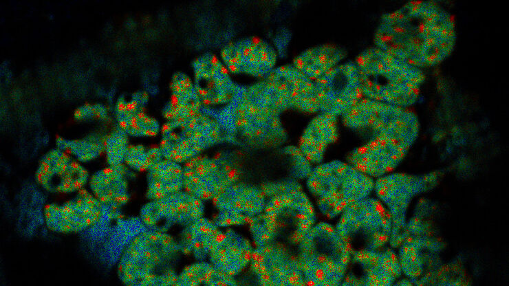
![[Translate to chinese:] Projection of a confocal z-stack. Sum159 cells, human breast cancer cells kindly provided by Ievgeniia Zagoriy, Mahamid Group, EMBL Heidelberg, Germany. Blue–Hoechst - indicates nuclei, Green–MitoTracker mitochondria, and red–Bodipy - lipid droplets](/fileadmin/_processed_/6/6/csm_Keyvisual-Cancer-cell-under-Cryo_Coral-Cryo_TechNote_999711acd8.jpg "[Translate to chinese:] Projection of a confocal z-stack. Sum159 cells, human breast cancer cells")
低温光学显微镜的新成像工具
荧光显微镜图像能够为cryo-FIB加工提供定位支持,其质量决定了所制备薄片的结果。本文描述了LIGHTNING技术是如何显著提高图像质量,以及如何利用该技术基于荧光寿命的信息来辨别样品的不同结构。
Loading...

如何对荧光结构三维定位以进行冷冻FIB切片
冷冻ET(电子断层扫描)是一种专用的透射电子显微镜技术,可以重建观察区域的三维体积。借助先进的冷冻EM(电子显微镜),图像分辨率可以提升到令人难以置信的亚纳米等级。因此,可以在细胞内的原生环境中研究蛋白质以及其他生物分子,从而揭示尚未探明的分子机制。由于细胞和组织必须薄到能够透过电子,样品必须进行切片以获取足够薄的样品体积(薄层)。为对样品中的靶区进行精确的三维定位,冷冻共聚焦显微镜是必不可少的工…
Loading...
![[Translate to chinese:] LNG-non-LNGHeLa cells labeled with light blue –Hoechst, Nuclei](/fileadmin/_processed_/c/6/csm_LNG-non-LNGHeLa-cells_828210c750.jpg "[Translate to chinese:] LNG-non-LNGHeLa cells. Cells kindly provided by I. Zagoriy, Mahamid Group, EMBL Heidelberg")
精确三维定位,实现EM成像——掌握精髓
低温电子断层扫描(CryoET)是一种成像技术,可以让研究人员以亚纳米分辨率观察蛋白质和其他大生物分子。了解分子的形状和结构,包括口袋和裂隙,可以帮助研究人员设计能够像拼图一样附着于分子的药物。低温ET成像也因此成为了解和治疗疾病和失调的重要基础。
Loading...

冷冻光电联用(Cryo-CLEM)之旅
本文主要介绍Cryo-CLEM技术及其为科学家带来的便益。此外,还特别说明了一些相关文献。
近期在冷冻电子显微镜工作流程领域取得的技术进步,让我们能够获取到细胞蛋白质社会学的3D数据,其分辨率更是达到前所未有的1纳米以下。工作流程中有一个步骤,需要从样品获取目标位置纳米级分辨率的图像,而要得到这样的结果,就需要用到冷冻光学显微镜。这种显微镜如果用于低温电子显微镜工作流程,通常就称为Cryo…
Loading...
![[Translate to chinese:] Cryo FIB lamella - Overlay of SEM and confocal fluorescence image. Target structure in yeast cells (nuclear pore proteine Nup159-Atg8-split Venus, red) marked by an arrow. Scale bar: 5 µm. Alegretti et al., Nature 586, 796-800 (2020).](/fileadmin/_processed_/c/d/csm_Targeting_Nuclear_Pore_Complexes_teaser_27b8f39513.jpg "[Translate to chinese:] Cryo FIB lamella - Overlay of SEM and confocal fluorescence image")
使用冷冻共聚焦显微镜定位活性循环核孔复合物
本文介绍了如何利用冷冻光学显微镜,尤其是冷冻共焦显微镜来提高冷冻工作流程的可靠性。评估了EM网格和样品的质量,并分析了目标结构的分布。本文展示了如何将冷冻共焦3D数据投射到SEM图像上,将感兴趣结构可靠地保留在FIB切割的薄片内,以便在冷冻TEM中进行进一步研究。
Loading...

Advancing Cell Biology with Cryo-Correlative Microscopy
Correlative light and electron microscopy (CLEM) advances biological discoveries by merging different microscopes and imaging modalities to study systems in 4D. Combining fluorescence microscopy with…
Loading...

改善冷冻电子断层扫描工作流程
徕卡显微系统有限公司和赛默飞世尔科技有限公司合作开发了一个整条技术路线的冷冻电子断层扫描工作流程。它确保从通过THUNDER成像仪EM冷冻CLEM(也可选择新版的CORAL Cryo冷冻共聚焦CLEM)预选与我们的EM GP2的玻璃化冷冻到Thermo Scientific Krios™ G3i Cryo TEM的3D图像重建的完全整合。所有仪器之间的无缝通信能够获得可靠的结果和可重现的实验。
