
Filter articles
标签
产品
![[Translate to chinese:] Murine esophageal organoids (DAPI, Integrin26-AF 488, SOX2-AF568) imaged with the THUNDER Imager 3D Cell Culture. Courtesy of Dr. F.T. Arroso Martins, Tamere University, Finland.](/fileadmin/_processed_/f/f/csm_THUNDER_Imager_3D_Cell_Culture_Murine-esophageal-organoid_LVCC_299fe0ce61.jpg "[Translate to chinese:] Murine esophageal organoids (DAPI, Integrin26-AF 488, SOX2-AF568) imaged with the THUNDER Imager 3D Cell Culture. Courtesy of Dr. F.T. Arroso Martins, Tamere University, Finland.")
Loading...
![[Translate to chinese:] Fluorescence microscopy image of liver tissue where DNA in the nuclei are stained with Feulgen-pararosanilin and visualized with transmitted green light.](/fileadmin/_processed_/0/6/csm_Fluorescence_microscopy_image_of_liver_tissue_0748f2a4d5.jpg "[Translate to chinese:] Fluorescence microscopy image of liver tissue where DNA in the nuclei are stained with Feulgen-pararosanilin and visualized with transmitted green light.")
落射荧光显微镜和反射对比显微镜
多年来,荧光显微镜一直仅使用透射光和暗场照明。随着时间的推移,对改进照明的需求不断增长,这导致了落射照明(也称为入射光照明)的发展。经过 40 年的发展和改进,落射照明荧光显微镜已成为生命科学、临床医学诊断和材料科学领域常规实验室工作和研究的实用方法。大部分开发工作由 Ploem 集团和 Leitz 公司(现为 Leica Microsystems)完成。
Loading...
![[Translate to chinese:] Molecular structure of the green fluorescent protein (GFP)](/fileadmin/_processed_/a/c/csm_Fluorescent-Proteins_5a50f9b8af.jpg "[Translate to chinese:] Molecular structure of the green fluorescent protein (GFP)")
荧光蛋白简介
本文概述了荧光蛋白及其光谱特性。随着 20 世纪 50 年代末荧光蛋白的发现,荧光显微技术发生了巨大变化。它始于 O. Shimomura 和来自水母(Aequorea victoria)的绿色荧光蛋白(GFP)[1]。后来出现了数百种 GFP…
Loading...
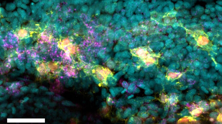
![[Translate to chinese:] Branched organoid growing in collagen where the Nuclei are labeled blue. To detect the mechanosignaling process, the YAP1 is labeled green.](/fileadmin/_processed_/a/e/csm_Branched_organoid_growing_in_collagen_dc289aa8c6.jpg "[Translate to chinese:] Branched organoid growing in collagen where the Nuclei are labeled blue. To detect the mechanosignaling process, the YAP1 is labeled green.")
检查癌症类器官的发展进程
德国慕尼黑工业大学的Andreas Bausch实验室研究细胞和生物体中不同结构和功能形成的细胞和生物物理机制。他的团队设计了新的策略、方法和分析工具,以量化微米和纳米等级的发展机制和动态过程。关键研究领域包括干细胞和类器官,从乳腺类器官到胰腺癌类器官,以更好地了解疾病模型。
Loading...

荧光入门介绍
荧光是George Gabriel Stokes于1852年首次报道的一种现象。他观察到萤石在紫外线照射后开始发光。荧光是光致发光的一种形式,是指一种材料被光照射后会发射出光子。发射光的波长比激发光更长。这种效应又称为斯托克斯位移。
Loading...
![[Translate to chinese:] Masson-Goldner staining of a hedgehog brain slice.](/fileadmin/_processed_/c/0/csm_Masson-Goldner_staining_of_hedgehog_brain_slice_a392b39de3.jpg "[Translate to chinese:] Masson-Goldner staining of a hedgehog brain slice.")
如何利用单个系统对组织学荧光样品进行成像
在本集MicaCame中,主持人Lynne Turnbull和Patric Pelzer将带您探寻生物样本染色的历史之旅。他们将解释为什么您通常必须选择为组织学样品或荧光样品选择特定的系统,以及如何利用新的成像技术克服这一点。
Loading...
如何从根本上简化成像设备的工作流程
本集MicaCam中,来自伦敦大学学院(UCL)的特邀嘉宾Christopher Thrasivoulou博士将从成像设备的角度讨论使用Mica的优势。他将讨论如何简化复杂生物系统的成像工作流程并实现自动化。这有助于科学家节省为获取有意义的量化分析结果而投入的时间和精力。为了举例说明此类工作流程,他还会展示如何对荧光标记的固定斑马鱼胚胎进行多色成像。
Loading...
![[Translate to chinese:]](/fileadmin/_processed_/0/f/csm_Unmixing_multicolour_widefield_fluorescence_images_teaser_56d2f9ce81.jpg "[Translate to chinese:]")
FLUOSYNC - 一种快速而温和的多色光谱拆分成像方法
在本白皮书中,我们重点介绍如何使用一种快速、可靠的方法在荧光显微镜下获得高质量多通道图像。FluoSync 将现有的光谱混合拆分方法与同步采集多个光谱探测范围相结合,一步到位。这样,多个荧光团可同时成像,而且无需担心荧光串扰、滤光片的选择或在高速成像下损失重要光子的问题。从样本中获得真正的信号从未如此容易。
Loading...
利用微流控技术保持活细胞成像期间的细胞健康
点播视频——在这集MicaCam中,我们将使用微流控技术探索对细胞形态的剪切应力,检查3D细胞培养期间营养物质补充对细胞生长的影响,并观察长期培养期间球状体的发育。
