Filter articles
标签
产品
Loading...

Mica: A Game-changer for Collaborative Research at Imperial College London
This interview highlights the transformative impact of Mica at Imperial College London. Scientists explain how Mica has been a game-changer, expanding research possibilities and facilitating…
Loading...

模式生物研究
模式生物是研究人员用来研究特定生物学过程的物种。 它们具有与人类相似的遗传特征,通常用于遗传学、发育生物学和神经科学等研究领域。 选择模式生物的原因通常是它们在实验室环境中易于保持和繁殖、生成周期短,或能够产生突变体来研究某些性状或疾病。
Loading...

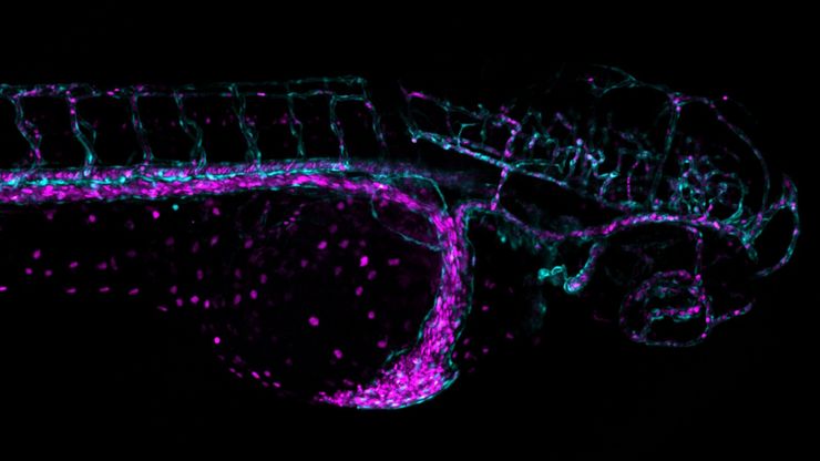
Overcoming Challenges with Microscopy when Imaging Moving Zebrafish Larvae
Zebrafish is a valuable model organism with many beneficial traits. However, imaging a full organism poses challenges as it is not stationary. Here, this case study shows how zebrafish larvae can be…
Loading...
![[Translate to chinese:] Salmonella biofilms 3D render [Translate to chinese:] Salmonella biofilms 3D render](/fileadmin/_processed_/9/6/csm_Salmonella_biofilms_3D_render_b444a820a0.jpg)
探索微生物世界:三维食品基质中的空间相互作用
Micalis 研究所是与 INRAE、AgroParisTech 和巴黎萨克雷大学合作的联合研究单位。其使命是开发食品微生物学领域的创新研究,以促进健康。在这一系列视频中,Micalis…
Loading...

Advancing Uterine Regenerative Therapies with Endometrial Organoids
Prof. Kang's group investigates important factors that determine the uterine microenvironment in which embryo insertion and pregnancy are successfully maintained. They are working to develop new…
Loading...
![[Translate to chinese:] The role of extracellular signalling mechanisms in the correct development of the human brain [Translate to chinese:] The role of extracellular signalling mechanisms in the correct development of the human brain](/fileadmin/_processed_/a/e/csm_The_role_of_extracellular_signalling_mechanisms_in_the_correct_development_of_the_human_brain_6b9e3b80f0.jpg)
在神经发育过程中,细胞是如何相互交流的?
细胞间通信是大脑发育过程中一个必不可少的过程,它受到多种因素的影响,包括细胞的形态、粘附分子、局部细胞外基质和分泌囊泡。在本次网络研讨会上,您将了解到对这些机制更深入的理解是如何推动对神经发育障碍的理解的。
Loading...

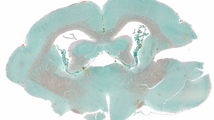
How to Streamline Your Histology Workflows
Streamline your histology workflows. The unique Fluosync detection method embedded into Mica enables high-res RGB color imaging in one shot.


