Filter articles
标签
产品
Loading...

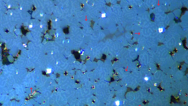
使用克尔显微镜快速可视化钢中的磁畴
磁性材料中磁畴与偏振光相互作用后光的旋转,称为克尔效应,使得使用克尔显微镜对磁化样品进行研究成为可能。它可以快速可视化材料表面的磁域。对于用于电气和电子设备的磁性材料(例如钢合金)的高效研发和质量控制,克尔显微镜可以发挥重要作用。本文详细描述了如何使用克尔显微镜对钢合金晶粒中的磁域进行成像。
Loading...

6英寸晶圆检测显微镜:可靠观察细微高度差异
本文介绍了一种配备自动化和可重复的DIC(微分干涉对比)成像的6英寸晶圆检测显微镜,无论用户的技能水平如何。制造集成电路(IC)芯片和半导体组件需要进行晶圆检测,以验证是否存在影响性能的缺陷。这种检测通常使用光学显微镜进行质量控制、故障分析和研发。为了有效地可视化晶圆上结构之间的小高度差异,可以使用DIC。
Loading...

术中OCT引导的青光眼支架修复手术
青光眼是导致全球不可逆失明的主要原因之一。小梁网切除术和导管分流引流术等历史悠久的手术技术会带来巨大的短期风险和潜在并发症。近年来,随着微创青光眼手术(MIGS)的出现,手术方法有了长足的发展,其特点是对组织的破坏最小、内路粘小管植入、手术时间短、器械简单、术后恢复快。
Loading...

Safe Wafer Loading for Microscope Inspection without Hand Contact
How automated silicon wafer loading for microscope inspection helps improve microelectronics process control and production efficiency is explained in this article. Manual handling of wafers has a…
Loading...

肿瘤重建外科的进展
在肿瘤重建外科的决策和患者护理方面,近年来发生了显著的变化。新的外科辅助技术正在帮助外科医生突破可实现的界限。这些技术包括:符合人体工程学的外科显微镜、吲哚菁绿(ICG)荧光、切割导向器和 3D 打印、增强现实以及高倍放大、全高清目镜图像注入,以及 2D 和 3D 视频录制。
Loading...

电池制造过程中的毛刺检测
毛刺是电池电极片边缘可能出现的缺陷,例如在制造过程中的分切环节。它们可能会因诸如短路等故障导致电池性能下降,并引发安全和可靠性问题。毛刺检测是电池生产质量控制的重要部分,对于生产具有可靠性能和寿命的电池至关重要。通过适当照明的光学显微镜可以在生产过程的关键步骤中快速可靠地对电极上的毛刺进行视觉检测。
Loading...

超薄切片技术电子书:定位、修块& 对刀
超薄切片技术正经历日新月异的发展,当今的显微镜系统对高质量切片、精准定位以及可重复的工作流程提出了更高要求。这本电子书整合了专家应用指南、自动化方法及实操指导,旨在帮助从初学者到资深镜检人员的每一位用户,在电镜、光电联用及体电镜工作流程中,获得一致且可靠的超薄切片。
Loading...

Flexibility and Efficiency in Minimally Invasive Spine Surgery
According to Prof. Alex Alfieri, Chief Physician and Head of clinic for Neurosurgery and Spinal surgery at the Cantonal Hospital Winterthur, Minimally invasive spine surgery (MISS) is transforming…
Loading...

High-Pressure Freezing for Organoids: Cryo CLEM & FIB Lift Out
Master cryo EM workflow steps for challenging 3D samples: when to choose HPF vs. plunge freezing, reproducible blotting/ice control, contamination aware transfers, Cryo CLEM 3D targeting in organoids,…

