
Filter articles
标签
产品
Loading...

From Bench to Beam: A Complete Correlative Cryo Light Microscopy Workflow
In the webinar entitled "A Multimodal Vitreous Crusade, a Cryo Correlative Workflow from Bench to Beam" a team of experts discusses the exciting world of correlative workflows for structural biology…
Loading...

Exploring the Structure and Life Cycle of Viruses
The SARS-CoV-2 outbreak started in late December 2019 and has since reached a global pandemic, leading to a worldwide battle against COVID-19. The ever-evolving electron microscopy methods offer a…
Loading...

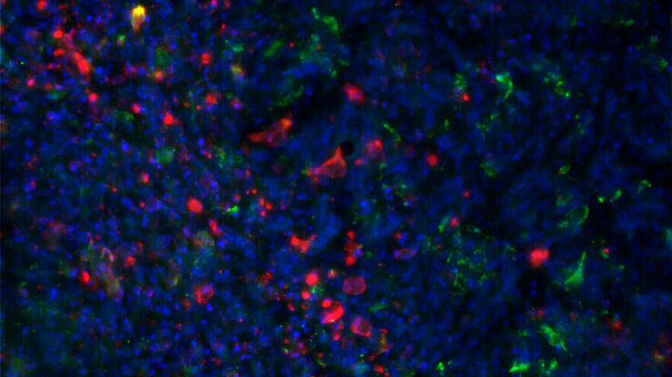
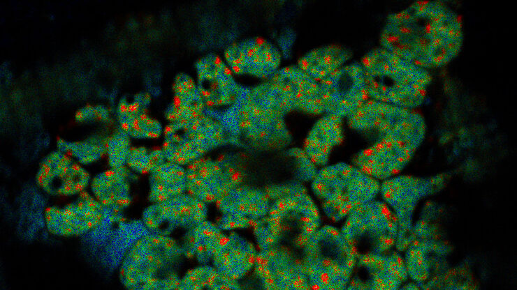
Advancing Cell Biology with Cryo-Correlative Microscopy
Correlative light and electron microscopy (CLEM) advances biological discoveries by merging different microscopes and imaging modalities to study systems in 4D. Combining fluorescence microscopy with…
Loading...

Workflows and Instrumentation for Cryo-electron Microscopy
Cryo-electron microscopy is an increasingly popular modality to study the structures of macromolecular complexes and has enabled numerous new insights in cell biology. In recent years, cryo-electron…
Loading...

改善冷冻电子断层扫描工作流程
徕卡显微系统有限公司和赛默飞世尔科技有限公司合作开发了一个整条技术路线的冷冻电子断层扫描工作流程。它确保从通过THUNDER成像仪EM冷冻CLEM(也可选择新版的CORAL Cryo冷冻共聚焦CLEM)预选与我们的EM GP2的玻璃化冷冻到Thermo Scientific Krios™ G3i Cryo TEM的3D图像重建的完全整合。所有仪器之间的无缝通信能够获得可靠的结果和可重现的实验。
Loading...
![[Translate to chinese:] Roland A. Fleck](/fileadmin/_processed_/6/1/csm_interview-fleck-teaser_9cfb9e0382.jpg "[Translate to chinese:] Roland A. Fleck")
专家在低温扫描电镜工作流程高压冷冻和冷冻断裂方面的知识
深入了解实验室工作方法并了解在EM样本制备过程中低温扫描电镜研究的优势。了解如何将高压冷冻、冷冻断裂和冷冻传送添加到低温扫描电镜工作流程中,以及徕卡组合如何确保这些不同步骤之间的兼容性。
Loading...
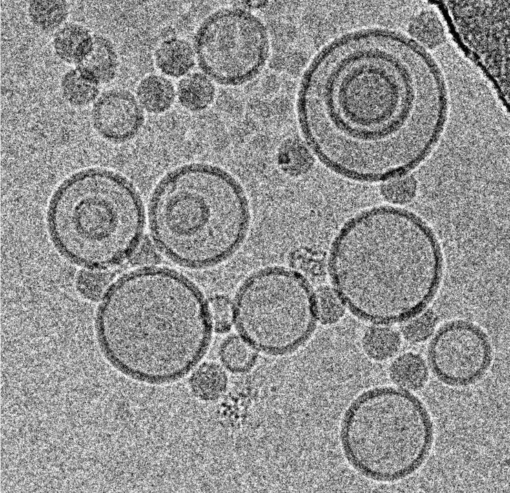
冷冻透射电子显微镜的投入式冷冻技术:应用
低温下观察完全含水、未染色样本的透射电子显微镜(cryo TEM)是结构生物学、细胞生物学、药理学和其他科学分支的通用工具。通过将标本放入冷冻剂中进行超快速冷冻(投入式冻结)是一种常用的方法,用于制备在透射电镜观察的各种标本。本文是对投入式冷冻的补充,介绍了在不同领域使用投入式冷冻标本的三种冷冻TEM应用。
