Filter articles
标签
产品
Loading...

Designing the Future with Stem Cell and RNA Technology
Visionary biotech start-up Uncommon Bio is tackling one of the world’s biggest health challenges: food sustainability. In this webinar, Stem Cell Scientist Samuel East will show how they use RNA…
Loading...

组织中的精密空间蛋白质组学信息
尽管可使用基于成像和质谱的方法进行空间蛋白质组学研究,但是图像与单细胞分辨率蛋白丰度测量值的关联仍然是个巨大的挑战。最近引入的一种方法,深层视觉蛋白质组学(DVP),将细胞表型的人工智能图像分析与自动化的单细胞或单核激光显微切割及超高灵敏度的质谱分析结合在了一起。DVP在保留空间背景的同时,将蛋白丰度与复杂的细胞或亚细胞表型关联在一起。
Loading...

采用徕卡THUNDER-DM6B观察SARS-CoV-2感染宿主细胞及其复制过程
冠状病毒2致重度急性呼吸综合征(SARS-CoV-2)
冠状病毒2致重度急性呼吸综合征(SARS-CoV-2)出现于2019年末,并快速传播全世界。由于其大面积的影响,研究人员对病毒的性质进行了深入的研究以期最终阻止大流行。一个重要的方面是病毒如何在宿主细胞中复制。Ogando及其同事的研究已经揭示了SARS-CoV-2的复制动力学、适应能力和细胞病理学。他们的工具之一是用荧光显微镜观察SARS…
Loading...
![[Translate to chinese:] Fluorescence microscopy image of liver tissue where DNA in the nuclei are stained with Feulgen-pararosanilin and visualized with transmitted green light. [Translate to chinese:] Fluorescence microscopy image of liver tissue where DNA in the nuclei are stained with Feulgen-pararosanilin and visualized with transmitted green light.](/fileadmin/_processed_/0/6/csm_Fluorescence_microscopy_image_of_liver_tissue_0748f2a4d5.jpg)
落射荧光显微镜和反射对比显微镜
多年来,荧光显微镜一直仅使用透射光和暗场照明。随着时间的推移,对改进照明的需求不断增长,这导致了落射照明(也称为入射光照明)的发展。经过 40 年的发展和改进,落射照明荧光显微镜已成为生命科学、临床医学诊断和材料科学领域常规实验室工作和研究的实用方法。大部分开发工作由 Ploem 集团和 Leitz 公司(现为 Leica Microsystems)完成。
Loading...
![[Translate to chinese:] Molecular structure of the green fluorescent protein (GFP) [Translate to chinese:] Molecular structure of the green fluorescent protein (GFP)](/fileadmin/_processed_/a/c/csm_Fluorescent-Proteins_5a50f9b8af.jpg)
荧光蛋白简介
本文概述了荧光蛋白及其光谱特性。随着 20 世纪 50 年代末荧光蛋白的发现,荧光显微技术发生了巨大变化。它始于 O. Shimomura 和来自水母(Aequorea victoria)的绿色荧光蛋白(GFP)[1]。后来出现了数百种 GFP…
Loading...

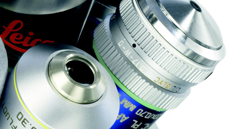
浸没式物镜
要用显微镜在高倍镜下检查试样标本,有许多因素需要考虑。其中包括分辨率、数字光圈数值孔径(NA)、物镜的工作距离以及物镜前透镜采集图像所用介质的折射率。在这篇文章中,我们将简要地看一下如何使用浸泡介质在盖玻片和物镜前透镜之间利用浸泡介质帮助提高NA和分辨率。此外,我们还将考虑空气和构成载玻片和、盖玻片的空气和玻璃的折射率,以及当光从一种介质传播到另一种介质时,如何使用浸没介质部分地减少失错配。此外,…
Loading...
![[Translate to chinese:] Images of smooth muscle cells during wound healing. Courtesy L.S. Shankman, Ph.D., University of Virginia. [Translate to chinese:] Images of smooth muscle cells during wound healing. Courtesy L.S. Shankman, Ph.D., University of Virginia.](/fileadmin/_processed_/4/3/csm_Smooth_muscle_cells_during__0df86436b1.jpg)
平滑肌细胞划痕愈合研究
本文主要讨论如何使用专门配置的徕卡倒置显微镜和台式细胞培养箱轻松、可靠地研究多孔板中培养的平滑肌细胞(SMC)的划痕愈合情况。血管受损后影响SMC增殖和迁移的信号转导情况在医学研究中有重要意义。由于SMC遍布全身,所以对其迁移情况的研究也有助于癌症和损伤的治疗。

![[Translate to chinese:] Neurons imaged with DIC contrast. [Translate to chinese:] Neurons imaged with DIC contrast.](/fileadmin/_processed_/3/1/csm_Neurons_imaged_in_DIC_8186d00fbe.jpg)
![[Translate to chinese:] Image of MDCK (Madin-Darby canine kidney) cells taken with phase contrast. [Translate to chinese:] Image of MDCK (Madin-Darby canine kidney) cells taken with phase contrast.](/fileadmin/_processed_/6/c/csm_MDCK_cells_in_phase_contrast_5821a63e2b.jpg)
