Filter articles
标签
产品
Loading...
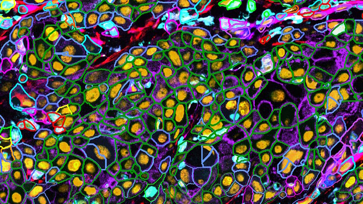
![[Translate to chinese:] Esophageal tissue with a squamous cell carcinoma labelled with the 4 biomarkers PanCk, DAPI, NaKATPase, and Vimentin. [Translate to chinese:] Esophageal tissue with a squamous cell carcinoma labelled with the 4 biomarkers PanCk, DAPI, NaKATPase, and Vimentin.](/fileadmin/_processed_/7/1/csm_Esophageal_Squamous_Cell_Carcinoma_4_Markers_7ac3f29e4b.jpg)
探索多重生物成像如何推进癌症研究
观看行业和学术专家进行的内容丰富的讨论,分享他们在研究中使用多重成像技术的知识。了解多重成像技术如何通过发现以前难以捉摸的分子洞察力,彻底改变肿瘤学、神经学和免疫学。利用先进的成像技术深入了解组织微环境,从而对代谢紊乱和癌症等疾病有新的认识。
Loading...

与卢克-加蒙(Luke Gammon)一起多重成像:推进您的空间生物学研究
多重成像是一种功能强大的技术,可让研究人员同时观察单个样本中的多个目标。这对于研究复杂的生物系统尤为重要,可以帮助研究人员更好地了解不同分子和途径之间是如何相互作用的。
Loading...
![[Translate to chinese:] PDAC Multiplexed imaging of CST panels enables an examination of immune cell components in pancreatic ductal adenocarcinoma (IPDAC) tissue on a single slide. [Translate to chinese:] PDAC Multiplexed imaging of CST panels enables an examination of immune cell components in pancreatic ductal adenocarcinoma (IPDAC) tissue on a single slide.](/fileadmin/_processed_/a/d/csm_Pancreatic_ductal_adenocarcinoma_tissue_d8790cf699.jpg)
表征肿瘤环境以揭示洞察和空间分辨率
肿瘤环境的表征可以为癌症进展和潜在治疗靶点提供更深入的见解。我们已经使用来自Cell Signaling Technology(CST)的各种IHC验证抗体,在胰腺癌的Cell DIVE研究中验证了30多种偶联抗体。
Loading...
![[Translate to chinese:] Pancreatic Ductal Adenocarcinoma with 5 biomarkers shown – SMA, PanCK PCK26, PanCK AE1, Vimentin, and Glut1. [Translate to chinese:] Pancreatic Ductal Adenocarcinoma with 5 biomarkers shown – SMA, PanCK PCK26, PanCK AE1, Vimentin, and Glut1.](/fileadmin/_processed_/3/a/csm_Pancreatic_Ductal_Adenocarcinoma_with_5_biomarkers_412898c37f.jpg)
借助多重成像深入了解胰腺癌的复杂性
胰腺癌是一种很难治疗的肿瘤疾病。Cell DIVE多重成像可以视觉呈现30种生物标志物以探测胰管癌的微环境。此面板可以检查肿瘤组织多个层级的问题,包括淋巴细胞、血管新生、转移、侵袭、炎症、缺氧、代谢和免疫。多重成像和分析可以对肿瘤组织中的许多生物活动提供更为深入的洞察信息,从而可以深入研究这些信息。
Loading...
![[Translate to chinese:] Pancreatic ductal adenocarcinoma tissue section imaged with Cell DIVE [Translate to chinese:] Pancreatic ductal adenocarcinoma tissue section imaged with Cell DIVE](/fileadmin/_processed_/f/8/csm_Pancreatic_ductal_adenocarcinoma_tissue_section_teaser_3e2f21e476.jpg)
多重成像的类型、优势和应用
与传统显微镜相比,多重成像技术能观察到更多的生物标记物,是一种新兴的、令人兴奋的从人体组织样本中提取信息的方法。通过同时观察多种生物标记物,可以协同探索以前只能单独探索的生物通路,并识别和探测复杂的组织和细胞表型。目前已有许多不同的多重成像方法,每种方法都采用不同的方法来实现更高的复杂性。


![[Translate to chinese:] Cell DIVE Multiplex Imaging Solution [Translate to chinese:] Cell DIVE Multiplex Imaging Solution](/fileadmin/_processed_/0/6/csm_Cell_DIVE_dad174e320.jpg)
