
Filter articles
标签
产品
Loading...

人工智能显微成像能够高效检测稀有事件
对稀有事件进行定位和选择性成像是许多生物样本研究过程的关键。然而,由于时间限制和高度的复杂性,有些实验无法做到,从而限制了获得新发现的前景。通过基于人工智能的显微成像检测稀有事件,这种工作流程将智能样本导航、图像采集工具和人工智能驱动的图像分析等不同功能融合起来共同协作,能够克服上述局限性。
Loading...
 stained to show the nucleus")
复杂3D数据集——人工智能赋能的空间数据分析
本期MicaCam为您提供切实的建议,教您从显微镜图像中提取可发表级别的分析结果。本期的特邀嘉宾来自徕卡显微系统的Luciano Lucas,他将为大家展示如何使用MICA的AI赋能软件进行图像分析。他将深度分析两张MICA的3D成像,探究不同可见生物元素之间的空间关系。本期的最后将会介绍如何创作高保真视频动画以及其他可用于发表文章的结果。
Loading...

人工智能显微图像分析-介绍
人工智能引领的显微图像分析和可视化是用于数据驱动型科学发现的一项强大工具。人工智能技术可以帮助研究人员应对具有挑战性的成像应用,让他们能够从图像中获取更多的信息。
Loading...
 and mito OM (red) in a live U2OS cell")
多色四维超分辨光片显微镜
人工智能显微术研讨会主要关注和讨论显微术和生物医学成像领域的最新人工智能技术和工具。在该科学演示中,Yuxuan Zhao展示了如何通过渐进式深度学习策略并结合“双环调制的SPIM”设计改善活细胞中的细胞器三维成像。
Loading...

精确分析宽视野荧光图像
利用荧光显微镜的特异性,即便是使用厚样品和大尺寸样品,研究人员也能够快速轻松地准确观察和分析生物学过程和结构。然而,离焦荧光会提高背景荧光,降低对比度,影响图像的精确分割。THUNDER 与Aivia 的组合可以有效解决这一问题。前者可以消除图像模糊,后者会使用人工智能技术自动分析宽视野图像,提高操作速度和精确性。下面,我们来详细了解下这一协作方法。
Loading...
人工智能驱动的像素分类器
通过人工操作获得可重复的结果需要具备专业知识,而且工作冗长乏味。但是,现在有一种方法可以克服这些挑战,通过加快这种分析来提取图像的真正价值并获得深入的认识。人工智能驱动的像素分类器可快速提供可重复的分割结果,克服了人工操作问题。与基于功能的传统自动化相比,它可以提供更可靠的结果。
Loading...
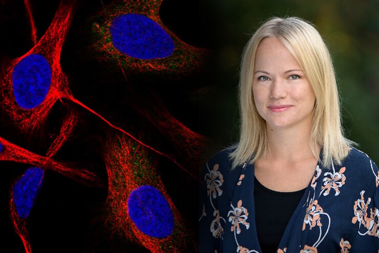
在显微成像和图像分析中运用人工智能和机器学习技术
Emma Lundberg 教授是瑞典 KTH 皇家理工学院细胞生物学蛋白质组学教授。她还是细胞图谱项目的总监,该项目是瑞典人类蛋白质图谱(HPA)项目不可或缺的组成部分,后者是用于研究人类蛋白质组的开源资源。细胞图谱项目是 HPA 的一部分,提供人类细胞系中 RNA 和蛋白质的表达及时空分布的高分辨率图像。Lundberg…
Loading...

在显微图像分析中运用机器学习技术
显微成像技术最近取得了令人振奋的进展,因此,在生物医学研究中采集的图像数据无论质量还是数量都呈指数级增长。但是,分析日益复杂的大型图像数据集以提取有意义的信息可能是一个既枯燥又耗时的过程,而且容易出现人为误差和偏差,这经常给许多研究人员造成生产效率瓶颈。
 enhanced with Aivia’s Pixel Classifier (right)")
