Filter articles
标签
产品
Loading...
![[Translate to chinese:] Pinhole diameter and diffraction pattern. [Translate to chinese:] Pinhole diameter and diffraction pattern.](/fileadmin/_processed_/9/f/csm_Pinhole_diameter_and_diffraction_pattern_fc8b6deecb.jpg)
共聚焦显微镜针孔效应
在操作共聚焦显微镜,或在讨论这种装置的特性和参数时,我们不可避免地提到针孔及其直径。这篇简短的文章是针对那些没有足够时间钻研共聚焦显微镜的理论和细节但又想了解针孔效应的用户们来解释针孔的意义。
Loading...

Rodent and Small-Animal Surgery
Learn how you can perform rodent (mouse, rat, hamster) and small-animal surgery efficiently with a microscope for developmental biology and medical research applications by reading this article.
Loading...

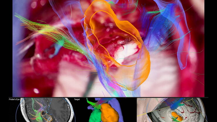
脑部导航
神经外科手术面临的挑战之一是在手术部位的定位。在切除肿瘤、去除动静脉畸形或夹闭动脉瘤时,外科医生常常需要在健康和功能性脑组织附近工作。在切除肿瘤时,挑战始终是尽可能保留健康组织。神经导航技术,也称为图像引导手术(IGS),使外科医生能够在切割之前规划理想的入路点,并通过提供术中定位来帮助执行该计划。
Loading...
![[Translate to chinese:] Left: Tissue cells marked with an immunolabel (FITC) illuminated with wide-band UV excitation. Note the tissue structure with blue autofluorescence. Right: Same tissue and same immunostaining with FITC label illuminated with epi-il [Translate to chinese:] Left: Tissue cells marked with an immunolabel (FITC) illuminated with wide-band UV excitation. Note the tissue structure with blue autofluorescence. Right: Same tissue and same immunostaining with FITC label illuminated with epi-illumination using narrow-band blue (490 nm) light. Note the increased image contrast (Ploem, 1967)](/fileadmin/_processed_/c/2/csm_Ploem_Figure_5_Autofluorescence_a_b_fbca553e26.png)
Milestones in Incident Light Fluorescence Microscopy
Since the middle of the last century, fluorescence microscopy developed into a bio scientific tool with one of the biggest impacts on our understanding of life. Watching cells and proteins with the…
Loading...
![[Translate to chinese:] HeLa cells stimulated with LPS. Image has been subjected to deconvolution. [Translate to chinese:] HeLa cells stimulated with LPS. Image has been subjected to deconvolution.](/fileadmin/_processed_/c/0/csm_HeLa_cells_stimulated_with_LPS_deconvolution_cf0b3cf706.jpg)
显微镜下的慢性炎症
在慢性炎症的过程中,身体的某些部位会反复发炎。许多人类疾病都是如此。在宽场光学显微镜的帮助下,可以对从细胞水平到整个生物体的潜在过程进行检查。本文介绍了几种宽场显微镜应用,如免疫荧光、活细胞成像、组织学和比率分析,以深入了解慢性炎症的发展、相关疾病及其治疗。
Loading...
![[Translate to chinese:] Wifi education solutions [Translate to chinese:] Wifi education solutions](/fileadmin/_processed_/c/e/csm_csm_wifi-education-solutions2_03_0a47178324_7705187c87.jpg)
选择学生显微镜需要考虑的因素
对于教师来说,选择教育显微镜并非易事。显微镜必须经得起并非总是小心翼翼的双手的日常使用,必须能够持续运行,还必须符合预算要求。尤其是学生用显微镜,实用性方面起着重要作用: 尺寸、重量、布线和设计在日常使用中非常重要,甚至在决定使用显微镜的设备和附件之前就应考虑到这一点。如果选择得当,教育显微镜将为大中小学的年轻人打开一扇通往微小细节的宇宙之窗,让他们对科学产生足够的兴趣,并将其作为自己的职业。
Loading...

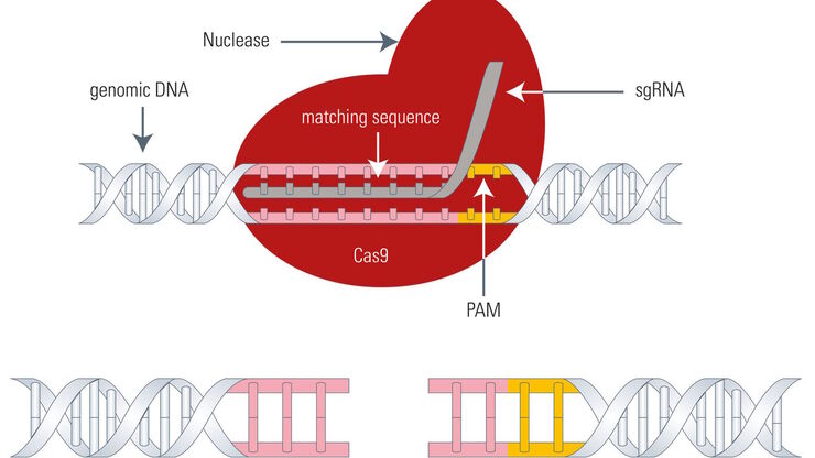
Gene Editing with CRISPR/Cas9 - Breakthrough in Genome Engineering
The CRISPR/Cas9 system is one of several different bacterial systems for defense against viral attacks. It consists of two main components. One is a small piece of RNA which binds to the viral target…
Loading...

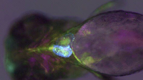
Imaging and Analyzing Zebrafish, Medaka, and Xenopus
Discover how to image and analyze zebrafish, medaka, and Xenopus frog model organisms efficiently with a microscope for developmental biology applications from this article.

