Filter articles
标签
产品
Loading...
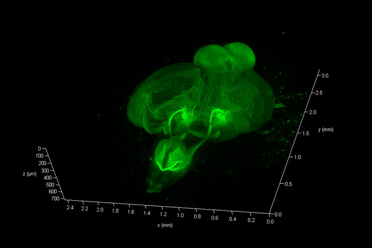
斑马鱼大脑高分辨率全器官成像
结构信息是理解复杂生物系统的关键,而脊椎动物的中枢神经系统是最复杂的生物结构之一。要想从发育中的斑马鱼身上分离出一个完整的大脑,我们需要覆盖大约10平方毫米的区域,深度在毫米范围内。通常,低倍透镜不能提供足够的分辨率来揭示神经组织中复杂结构之间的相互作用。此外,由于散射过程,使用共聚焦显微镜在致密生物组织内成像深度通常限制在大约10微米。
Loading...
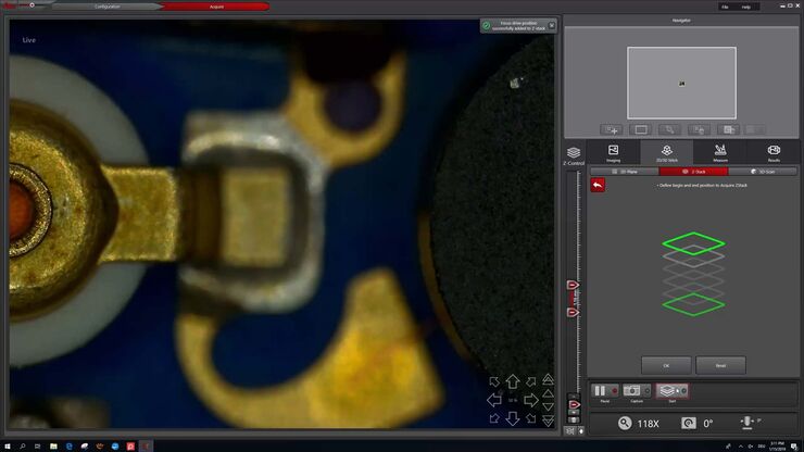
如何快速进行Z堆栈
为您的2D和3D分析节省时间。观看这个视频了解全新用户界面,专为DVM6数码显微镜开发的LAS X.next,这个视频演示了如何用几下点击来做一个快速的Z-Stack。
Loading...
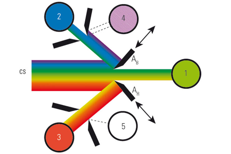
What is a Spectral Detector (SP Detector)?
The SP detector from Leica Microsystems denotes a compound detection unit for point scanning microscopes, in particular confocal microscopes. The SP detector splits light into up to 5 spectral bands.…
Loading...
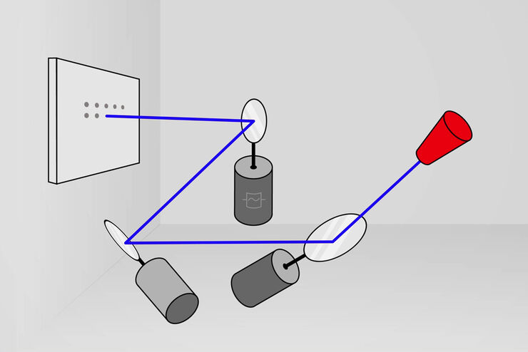
什么是共振扫描头?
共振扫描头是一种振镜扫描系统,可使用于单点扫描显微镜(真共焦和多光子激光扫描)快速获取图像。为了跟踪快速的过程,特别是在活体样品中,需要较高的采集速度。并且提供更好的荧光信号,减少光漂白 (1)。
Loading...
![[Translate to chinese:] Elucidate cancer development on sub-cellular level by in-vivo like tumor spheroid models. [Translate to chinese:] Elucidate cancer development on sub-cellular level by in-vivo like tumor spheroid models.](/fileadmin/_processed_/f/5/csm_3d-biology-workflow-DLS_5efeff5312.jpg)
利用光片显微镜改进三维细胞生物学工作流程
了解癌症发生过程中的亚细胞机制对于癌症治疗至关重要。常见的细胞模型涉及作为单层生长的癌细胞。然而,这种方法忽视了肿瘤细胞与其周围微环境之间的三维相互作用。为了贴近自然环境理解恶性肿瘤的发展和进程,对癌症微环境的详细表征至关重要。
Loading...
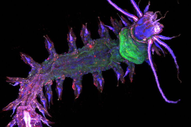
什么是视场扫描仪?
视场扫描器是单点共聚焦显微镜中使用的振镜扫描组件,可对大视场尺寸进行正确的光学记录。与传统的双镜扫描仪相比,视场扫描仪采用了三镜概念,在不影响高速扫描的情况下提供了更高的照明均匀性。振镜扫描仪的速度和位置可控,允许缩放和平移功能以及调整扫描频率。它们还可以进行静止点照明,例如 FCS 测量所需的照明。
Loading...
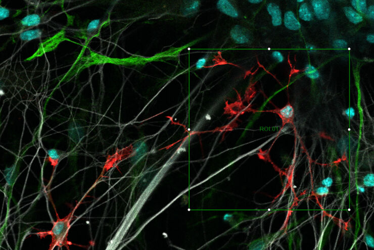
解析视场数(RFN)
光学显微镜的视场数(FN)表示视野大小(FOV)。它对应于中间图像中通过目镜可以观察到的区域。虽然我们不能一次观测到很大的视野,但人眼可以扫描并整合整个视野的结构特征。重要的是,该领域的大小和分辨率适合人眼能力。
Loading...
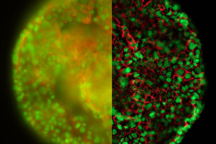
清晰对比、无雾的 3D 样本实时图像
历史上,宽场显微镜并不适合对大样本/标本体积进行成像。图像背景(BG)主要来源于观察样本的失焦区域,显著降低了成像系统的对比度、有效动态范围和最大可能的信噪比(SNR)。记录的图像显示出典型的雾霭,并且在许多情况下,无法提供进一步分析所需的细节水平。处理厚三维样本的研究人员要么使用替代显微镜方法,要么尝试通过后处理一系列图像来减少雾霭。
Loading...
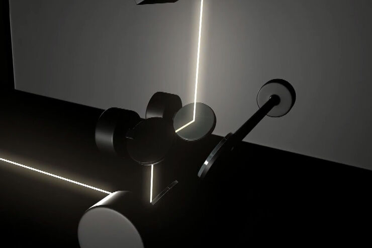
什么是串联扫描器?
串联扫描器集成了两种不同类型的扫描技术于一体,以实现真正的共焦点扫描。该系统包括一个三镜头扫描基座,其x轴扫描器可以与一个电动装置进行互换。这一组合不仅允许通过FOV扫描器实现大面积高分辨率扫描,还可以通过共振扫描器对极速过程进行扫描,两者均在同一仪器中完成。

