
Filter articles
标签
产品
Loading...

显微牙科:可视化和舒适性
在牙科手术过程中,显微镜不仅能提供更好的视觉效果,还有助于确保体位正确,避免背痛和颈部损伤。来自沙特阿拉伯利雅得市萨法德牙科中心的Dr.Iyad Ghoneim在中东迪拜国际口腔医学展览会牙科大会的演讲中分享了他使用Leica PROvido外科手术显微镜的经验。
Loading...
THUNDER成像:高效、灵活、易操作,让您的日常成像工作流更轻松
本次网络研讨会将展示 THUNDER 在许多不同生命科学应用中的多功能性和性能:从计数视网膜切片中的细胞核和癌组织切片中的 RNA 分子,到监测阿拉伯芥幼苗中的钙波等等。
Loading...
 from the desired fluorescent signal from the cell membrane (green).")
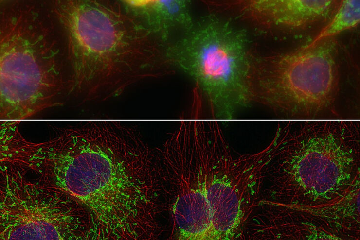
学习如何从共聚焦图像中去除自发荧光
了解自发荧光的常见原因以及如何将其从共聚焦显微镜图像中去除。根据应用的不同,自发荧光的来源可能有很多种,但幸运的是,同样也有很多的解决方案--从更换介质到使用荧光寿命成像和近红外染料。
Loading...
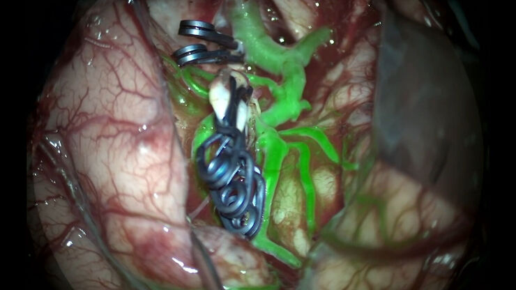
GLOW800增强现实荧光技术在动脉瘤治疗中的应用
Feres Chaddad博士教授的这个案例研究讨论了通过显微外科夹闭术治疗未破裂的MCA(大脑中动脉)和PCOM(后交通动脉)动脉瘤。这说明了增强现实荧光GLOW800借助实时血管血流增强技术,对大脑解剖结构获取增强实时视图,在动脉瘤夹闭前后为外科医生提供帮助。
Loading...

偏振光显微镜影像图集
偏振光显微镜(又称为偏光显微镜)是一种应用于不同领域的重要方法,包括研究和质量保证。它不仅仅是在高倍率和高分辨率下产生图像,这通常是用普通光学显微镜完成的。
通过检查样本的形状、结构、颜色、双折射和进一步的光学性质,可以获得有关样本结构、光学性质和成分的附加信息。
Loading...

金相学 – 介绍
本文概述了金相学和金属合金的特征分析。合金微观结构的研究使用到不同的显微观察技术,即晶粒、相、夹杂物等的微观结构。金相学是从了解合金微观组织对宏观性能影响发展而来的一门学科。所获得的知识可用于合金材料的设计、开发和制造。
Loading...
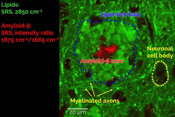
Stimulated Raman Scattering Microscopy Probes Neurodegenerative Disease
Despite decades of research, the molecular mechanisms underlying some of the most severe neurodegenerative diseases, such as Alzheimer’s or Parkinson’s, remain poorly understood. The progression of…
Loading...
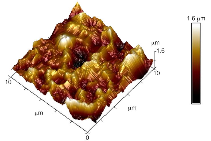
表面计量学简介
本报告简要讨论了几种常用于评估表面形貌(也称为表面纹理或表面光洁度)的重要计量技术和标准定义。随着纳米技术、薄涂层以及电路和装置小型化的出现,表面计量学已成为一个极其重要的科学和工程领域。
