Filter articles
标签
产品
Loading...

如何提高微电子元件检测性能
在检查硅片或微机电系统时,你需要看更多吗?你想得到与电子显微镜相似的清晰详细的样本图像吗?
观看这个免费的网络研讨会,了解更多关于强大的成像和对比技术,可以提高您的检查性能。您将了解如何克服分辨率标准,而不需要浸油或转移到SEM,以实现您想要的检测结果。
Loading...

荧光显微镜如何为工业应用带来益处
观看这个免费的网络研讨会,了解更多关于荧光显微镜在工业应用中的用途。我们将涵盖一系列调查研究,在这些研究中,荧光对比度为样本属性提供了新的见解,例如纤维、文件、涂料、建筑材料、电子产品和食品的属性。您将看到使用荧光有多么简单,同时还将了解样本制备和潜在局限性。
Loading...

How to Select a Microscope for Cataract Surgery
What to consider in the selection of an ophthalmic microscope for cataract procedures. Bearing these aspects in mind will equip surgeons well for talks with manufacturer representatives. Many…
Loading...

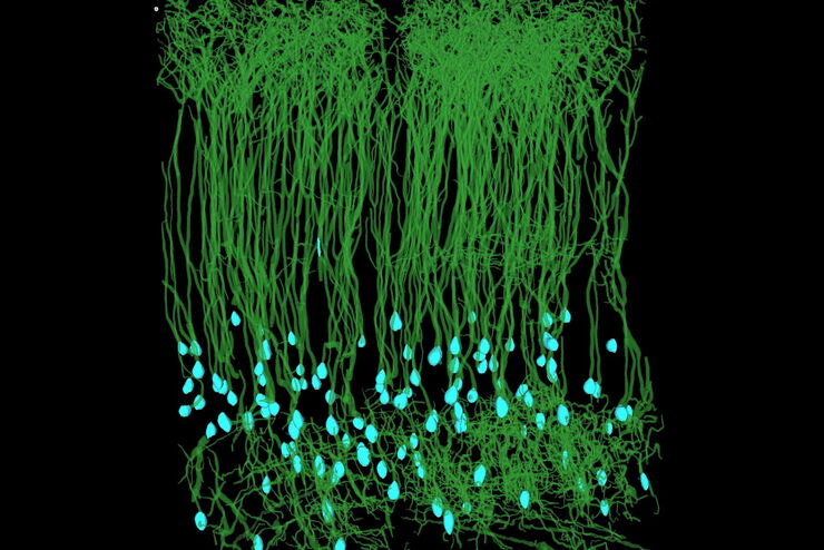
自动化加速神经元图像分析
复杂神经投射的检测能力主要取决于大规模神经元网络的精确重建。神经科学研究中的大多数数据析取方法都非常耗时和易错,进而导致进度延误和错误。在本次研讨会中,Aivia将演示如何利用自动化技术提升图像分析工作流的效率
Loading...

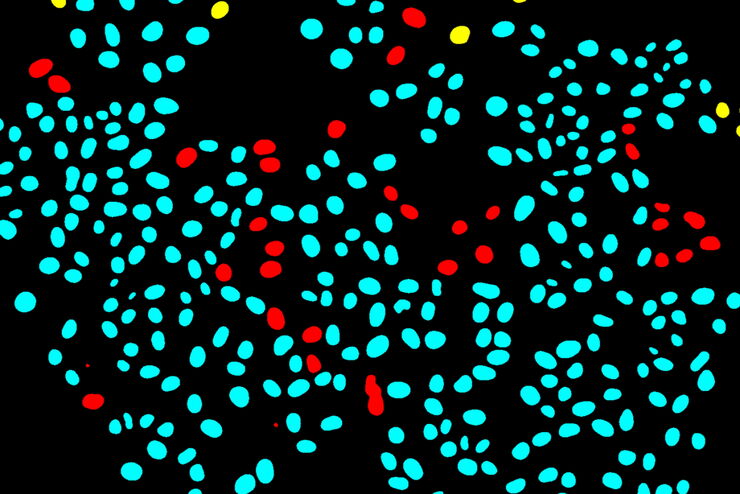
使用深度学习技术追踪单细胞
人工智能解决方案在显微镜领域的应用不断拓展。从自动化目标分类到虚拟染色,机器学习和深度学习技术在帮助显微镜学家简化分析工作的同时,也在持续推动科学技术领域的突破。


![[Translate to chinese:] Routine inspection microscope Ivesta 3 [Translate to chinese:] Routine inspection microscope Ivesta 3](/fileadmin/_processed_/a/a/csm_Ivesta_3_integrated_monitor_users_2_dc18a68be8.jpg)

