Filter articles
标签
产品
Loading...

福斯特共振能量转移 (FRET)
荧光描述的是分子或原子在通过光的吸收激发电子系统后,自发发射光子的过程。发射的光子通常能量较低,因此波长较长(斯托克斯位移)。例如,蓝光激发可能导致绿色发射。如果第二个荧光分子能够吸收绿色光子,则该分子的发射再次发生斯托克斯位移,例如变为红色。这种再吸收在浓密样品中会导致测量误差(部分“内滤”效应)。在低浓度样品中,再吸收非常罕见。
Loading...
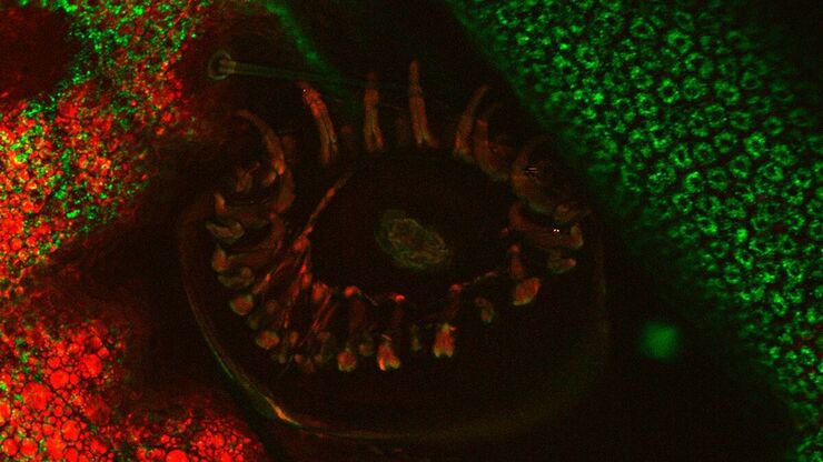
CARS 相干反斯托克斯拉曼散射显微镜简介
共聚焦和多光子成像技术仍然是对生物样本进行复杂研究的首选方法。这些技术可将生物样本中的典型结构或动态过程可视化,并依赖于样本中现有的自发荧光物质或合适的荧光染料。传统染色方法的缺点显而易见:标记耗时,染料会随时间褪色。此外,染料会失去强度并改变样本。染料通常会产生光毒性,对样本造成伤害,进而影响实验结果。CARS(相干反斯托克斯拉曼散射)显微镜是一种无需染料的方法,它通过显示结构分子的内在振动对比…
Loading...
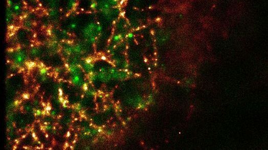
超分辨率 GSDIM 显微镜
纳米级技术GSDIM(基态耗尽显微镜后单分子返回)提供了细胞内蛋白质和其他生物分子空间排列的详细图像。目前市场上已有首个商业系统(Leica SR GSD),它正在帮助将GSDIM技术推广给更多研究实验室和成像中心的用户。
Loading...
![[Translate to chinese:] Scheme of a 2D mosaic scan. Drosophila melanogaster (eye section) [Translate to chinese:] Scheme of a 2D mosaic scan. Drosophila melanogaster (eye section)](/fileadmin/_processed_/e/b/csm_Mosaic_Intro_04_c164a21165.jpg)
马赛克图片
共焦激光扫描显微镜被广泛用于生成细胞、亚细胞结构甚至单分子的高分辨率三维图像。不过,越来越多的科学家正将生物研究的重点从单细胞研究扩展到整个器官和生物体,分析动物体内复杂的相互作用。
Loading...
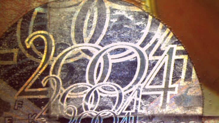
Is that Document Genuine or Fake? How do They Identify Fake Documents?
This article shows how forensic experts use microscopy for analysis to identify counterfeit, fake documents, such as ID cards, passports, visas, certificates, etc. Then they know if it is genuine or…
Loading...
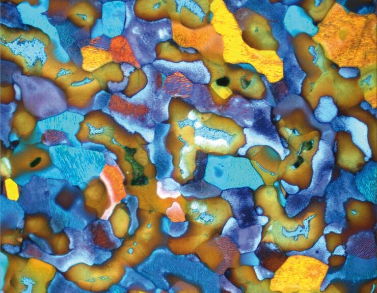
金相学:色彩与相衬度分析- 微观结构对比分析
显微组织形貌的检测在材料科学和失效分析中起着决定性的作用。在光学显微镜中有许多可能使材料的真实结构可视化。本文中显示的图像示例演示了使用的这些技术的信息及潜力。
Loading...

共聚焦光学截面厚度
共聚焦显微镜用于光学切片比较厚的样本。最直接的问题是:1. 什么是“比较厚的样本”;2. 光学切片到底有多厚?这两个问题在一定程度上是相关的,即假设一份厚样本至少比光学切片厚10倍。
Loading...
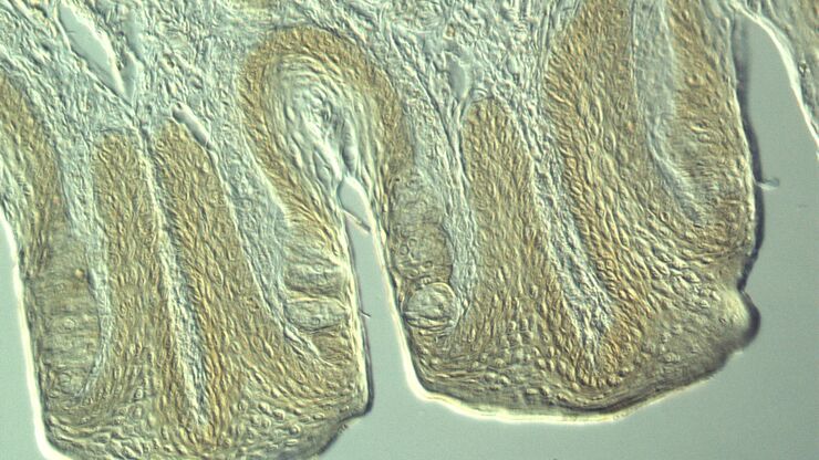
Optical Contrast Methods
Optical contrast methods give the potential to easily examine living and colorless specimens. Different microscopic techniques aim to change phase shifts caused by the interaction of light with the…
Loading...
![[Translate to chinese:] Modulation contrast visualizes transparent, low-contrast specimens. [Translate to chinese:] Modulation contrast visualizes transparent, low-contrast specimens.](/fileadmin/_processed_/9/d/csm_Modulation_contrast_visualizes_transparent_low-contrast_specimens_8a65b12740.jpg)
集成调制对比度 (IMC)
霍夫曼调制对比已成为观察未染色、低对比生物标本的标准。其创新的技术实现大大简化了操作,提高了使用的灵活性。将调制器集成到现代倒置显微镜的光束路径中,就可以使用各种明视野或相位物镜,而无需选择少量专用物镜。

