
Filter articles
标签
产品
Loading...

组织中的精密空间蛋白质组学信息
尽管可使用基于成像和质谱的方法进行空间蛋白质组学研究,但是图像与单细胞分辨率蛋白丰度测量值的关联仍然是个巨大的挑战。最近引入的一种方法,深层视觉蛋白质组学(DVP),将细胞表型的人工智能图像分析与自动化的单细胞或单核激光显微切割及超高灵敏度的质谱分析结合在了一起。DVP在保留空间背景的同时,将蛋白丰度与复杂的细胞或亚细胞表型关联在一起。
Loading...
![[Translate to chinese:] These images show the microstructure of a hard metal with 10% cobalt which is used for heavy-duty tools. The large increase in magnification of the right image (compared to the left) has a risk of being outside the useful range or, in other words, empty magnification.](/fileadmin/_processed_/d/b/csm_Microstructure_of_a_hard_metal_which_is_used_for_heavy-duty_tools_69233d4576.jpg "[Translate to chinese:] Microstructure of a hard metal with 10% cobalt which is used for heavy-duty tools.")
当心“无效”放大率
在一个最简化的情况,光学显微镜由一个靠近标本的透镜(物镜)和一个靠近眼睛的透镜(目镜)组成。显微镜放大率是两个显微镜透镜系数的乘积。比如,40倍物镜和10倍目镜可以得到400倍放大率。
Loading...

How to Study Gene Regulatory Networks in Embryonic Development
Join Dr. Andrea Boni by attending this on-demand webinar to explore how light-sheet microscopy revolutionizes developmental biology. This advanced imaging technique allows for high-speed, volumetric…
Loading...
![[Translate to chinese:] Multiplexed Cell DIVE imaging of Adult Human Alzheimer’s brain tissue section demonstrating expression of markers specific to astrocytes (GFAP, S100B), microglia (TMEM119, IBA1), AD-associated markers (p-Tau217, β-amyloid) and immune cells such as CD11b+, CD163+, CD4+, and HLA-DRA+, clustered around the β-amyloid plaques.](/fileadmin/_processed_/b/b/csm_Alzheimers_brain_tissue_section_showing_astrocytes_microglia_immune_cells_6f24036a9f.jpg "[Translate to chinese:] Multiplexed Cell DIVE imaging of Adult Human Alzheimer’s brain tissue section demonstrating expression of markers specific to astrocytes , microglia , AD-associated markers and immune cells clustered around the β-amyloid plaques")
Spatial Analysis of Neuroimmune Interactions in Alzheimer’s Disease
Alzheimer’s disease (AD) is a complex neurodegenerative disorder characterized by neurofibrillary tangles, β-amyloid plaques, and neuroinflammation. These dysfunctions trigger or are exacerbated by…
Loading...
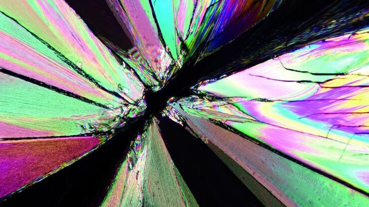
偏振光显微观察
偏光显微镜通常应用于材料科学和地质学领域,根据矿物的折射特性和颜色来识别矿物。在生物学中,偏光显微镜通常用于晶体等双折射结构的识别或成像,或用于植物细胞壁中纤维素和淀粉粒的成像。
Loading...

A Guide to Spatial Biology
What is spatial biology, and how can researchers leverage its tools to meet the growing demands of biological questions in the post-omics era? This article provides a brief overview of spatial biology…
Loading...

An Introduction to Laser Microdissection
The heterogeneity of histological and biological specimens often requires isolation of specific single cells or cell groups from surrounding tissue before molecular biology analysis can be carried…
Loading...
![[Translate to chinese:] GLP-1 and PYY localized to distinct secretory pools in L-cells.](/fileadmin/_processed_/0/2/csm_L-cells_366ce08e69.jpg "[Translate to chinese:] GLP-1 and PYY localized to distinct secretory pools in L-cells.")
前沿成像技术用于 GPCR 信号传导
通过这个按需网络研讨会,提升您的药理研究,了解 GPCR 信号传导,并探索旨在理解 GPCR 信号如何转化为细胞和生理反应的尖端成像技术。发现领先的研究,扩展我们对这些关键通路的认识,以寻找新的药物发现途径。
Loading...

Revealing Neuronal Migration’s Molecular Secrets
Different approaches can be used to investigate neuronal migration to their niche in the developing brain. In this webinar, experts from The University of Oxford present the microscopy tools and…
