Filter articles
标签
产品
Loading...
![[Translate to chinese:] Particles which could be found during cleanliness analysis of parts and components. [Translate to chinese:] Particles which could be found during cleanliness analysis of parts and components.](/fileadmin/_processed_/8/f/csm_Particles_found_during_cleanliness_analysis_9909ee7c63.jpg)
汽车零部件的清洁度
本文讨论了ISO 16232标准和VDA 19指南,并简要总结了颗粒物分析方法。它们为汽车零部件在微粒污染方面的清洁度提供了重要标准。此类颗粒物会对产品性能和寿命产生影响。在清洁度分析中,可以使用自动光学显微镜方法来确定颗粒物类型、大小和造成损坏的可能性。有时,需要更多成分信息,才能准确找到潜在的损害和污染源。这时候就需要借助激光光谱(LIBS)或电子显微镜。
Loading...
![[Translate to chinese:] GLOW800 Augmented Reality fluorescence in Moyamoya disease treatment. Image courtesy of Prof. Dr. Feres Chaddad [Translate to chinese:] GLOW800 Augmented Reality fluorescence in Moyamoya disease treatment](/fileadmin/_processed_/b/b/csm_GLOW800_Augmented_Reality_Moyamoya_disease_treatment_cc855dece6.jpg)
AR如何帮助烟雾病的手术治疗
烟雾病是一种罕见的慢性闭塞性脑血管疾病,其特点是颈内动脉末端进行性狭窄,大脑底部有异常的血管网络。搭桥手术是烟雾病的一种外科治疗方式。以下案例研究中,Feres Chaddad教授解释了GLOW800增强现实荧光如何支持此类手术,并详细介绍了一名35岁患者的手术治疗步骤。
Loading...

用MICA完成Caspase 3/7多色检测
Caspases与细胞凋亡过程相关,因此可以利用caspase检测来确定细胞是否正在经历这种程序化的细胞死亡。这些检测可以通过例如流式细胞仪、平板读数仪实现,也可以在显微镜上完成,显微镜可为量化数据补充可见的结构信息。在这篇文章中,我们描述了MICA是如何用于caspase 3/7测定。借助Navigator或像素分类器等工具,MICA让设置、执行和分析caspase…
Loading...

如何获得具有完全时空相关性的多标记实验数据
首期MicaCam会聚焦于活细胞实验当中的挑战。我们的主持人Lynne Turnbull和Oliver Schlicker将以活细胞内线粒体活动研究为例,手把手为您展示如何用多孔板培养箱设计您的实验,以及如何分析结果。
Loading...

使用口腔显微镜改善人体工程学
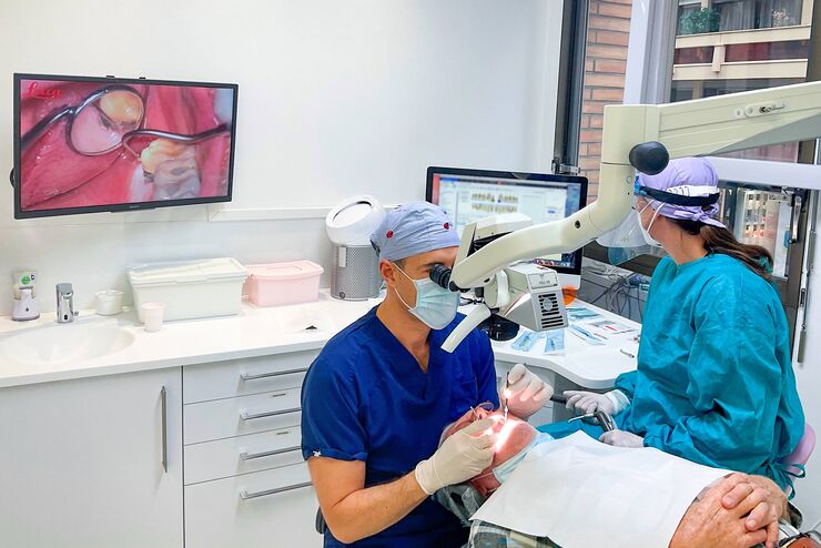
在这里,我们将向您展示牙科外科医生兼人体工程学顾问David Blanc医生如何使用带有超低角度双目镜筒的口腔显微镜提高身体舒适度。通过优化人体工程学设计,Blanc医生能够在为患者进行高精度显微牙科治疗时避免颈部弯曲。
Loading...
![[Translate to chinese:] Urethra and phalloplasty anastomosis [Translate to chinese:] Clinical case: radial forearm free flap preparation with prelaminated urethra and phalloplasty anastomosis](/fileadmin/_processed_/b/e/csm_Urethra_and_phalloplasty_anastomosis_78aad57c5b.jpg)
增强现实荧光技术在桡侧前臂皮瓣游离阴茎成形术中的应用
这个手术中,首席显微外科医生教授及其团队进行了桡侧前臂游离皮瓣阴茎成形术,并使用ICG荧光成像来显示整个皮瓣中从吻合口到阴茎远端的血流。教授展示了增强现实荧光技术除了常见的临床症状外,还能提供检查血流的状态。
Loading...
![[Translate to chinese:] Identification of distinct structures_roundworm_Ascaris_female [Translate to chinese:] Identification of distinct structures_roundworm_Ascaris_female](/fileadmin/_processed_/a/b/csm_Identification_of_distinct_structures_roundworm_Ascaris_female_eddbe9bbff.jpg)
从概览中查找相关样本细节
在从图像到图像的搜索中切换到快速查看整个样本概览,并即刻识别重要的样本细节。利用这些知识,使用载玻片、培养皿和多孔板的模板自动设置高分辨率图像采集。LAS X Navigator软件像是样本细胞的GPS,总能为用户指明通向高质量数据的清晰路径,这是生命科学平台STELLARIS和THUNDER成像仪上的一款强大的导航工具。LAS X Navigator支持将宽场、立体或共聚焦实验与舞台应用相结合。


![[Translate to chinese:] Developing zebrafish (Danio rerio) embryo, from sphere stage to somite stages. [Translate to chinese:] Developing zebrafish (Danio rerio) embryo, from sphere stage to somite stages.](/fileadmin/_processed_/8/f/csm_Developing_Zebrafish_embryo_MicaCam_teaser_81cef69cf1.jpg)
